Anti-Histone [F152C25W] Universal Antibody
![Anti-Histone [F152C25W] Universal Antibody](https://www.kerafast.com/MediaStorage/Product/Images/Large/1152_2001202001263611510.jpg "Anti-Histone [F152C25W] Universal Antibody")
This mouse IgG2a monoclonal antibody [F152C25W] was generated from an autoimmune mouse model and recognizes bovine, hamster, human, mouse and marsupial histones (all classes; H1 and core histones).
Highlights:
- Reacts with bovine, hamster, human, mouse and marsupial histones assayed (all classes; H1 and core histones)
- Suitable for Western Blot, ELISA and Immunofluorescence applications
Histones are the chief organizing proteins of DNA in the nucleus of all eukaryotic cells. They can be grouped into five major classes, whereof four form a nucleosome core particle that DNA is wrapped around, and the fifth organizes the linker DNA between the nucleosomes. Histones are among the most highly conserved proteins between species, which underscores their essential function for life of eukaryotic cells.
From the laboratory of Kim S. Wise, PhD, University of Missouri - Columbia.
 Part of The Investigator's Annexe program.
This mouse IgG2a monoclonal antibody [F152C25W] was generated from an autoimmune mouse model and recognizes bovine, hamster, human, mouse and marsupial histones (all classes; H1 and core histones).
Highlights:
- Reacts with bovine, hamster, human, mouse and marsupial histones assayed (all classes; H1 and core histones)
- Suitable for Western Blot, ELISA and Immunofluorescence applications
Histones are the chief organizing proteins of DNA in the nucleus of all eukaryotic cells. They can be grouped into five major classes, whereof four form a nucleosome core particle that DNA is wrapped around, and the fifth organizes the linker DNA between the nucleosomes. Histones are among the most highly conserved proteins between species, which underscores their essential function for life of eukaryotic cells.
From the laboratory of Kim S. Wise, PhD, University of Missouri - Columbia.
![]() Part of The Investigator's Annexe program.
Part of The Investigator's Annexe program.
| Product Type: | Antibody |
| Antigen: | Histones (all classes; H1 and core histones) |
| Molecular Weight: | Various, per known histone species |
| Isotype: | IgG2a |
| Clonality: | Monoclonal |
| Clone Name: | F152C25W |
| Reactivity: | All vertebrate species assayed: bovine, hamster, human, mouse and marsupial |
| Immunogen: | Naturally occurring autoantibody |
| Species Immunized: | Mouse |
| Purification Method: | Protein G |
| Buffer: | 0.1M Sodium Phosphate, pH 7.4, 0.15M NaCl, 0.05% (w/v) Sodium Azide |
| Tested Applications: | WB (1:1000), IF (1:1000 ), ELISA |
| Storage: | -20C |
| Shipped: | Cold packs |
Western Blot
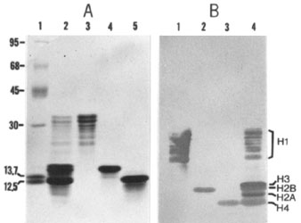
(Panel A) Proteins separated by SDS-PAGE (Laemmli system, 10-18% linear gradient) were stained with Coomassie Blue. Samples include:standard mol wt markers (lane 1), expressed in kilodaltons at left; histone mixture (lane 2); histone H1 (lane 3); histone H2B (lane 4), and histone H4 (lane 5). Fractionated or purified histones from calf thymus were obtained from Boehringer Mannheim Biochemicals. (Panel B) Western immunoblot of proteins separated by SDS-PAGE and stained with mAb F152C25W. Blots were generated from a separate gel (run under the same conditions as in panel A) containing 5 ug per well of: histone H1 (lane 1); histone H2B (lane 2); histone H4 (lane 3), and histone mixture (lane 4). The positions of the various histones are indicated.
Procedure adapted from: Hardin, JA. et al., Proc. Natl. Acad. Sci. USA 80:7410-7414.
Immunofluorescence Microscopy
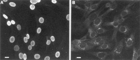
Fibroblast monolayers were fixed in cold methanol and incubated with mAb followed by FITC-conjugated goat anti-mouse Ig secondary Ab. (Bars = 10 microns). (Panel A): Incubated with mAb F152C25W; shows smooth, intense, specific nuclear staining. (Panel B): Incubated with irrelevant mAb of different specificity; to emphasize unstained nuclei.
If you publish research with this product, please let us know so we can cite your paper.