NO-Bio Sulfinic Acid Probe

NO-Bio is a chemoselective probe for detecting sulfinylated proteins in cell lysates.
Highlights:
- Rapid, selective and general technology for sulfinic acid detection in biological samples
- Consists of 2-nitroso terephthalic acid warhead coupled to a biotin tag by a PEG spacer
- 2-nitroso terephthalic acid phenyl ester moiety forms stable adducts with protein sulfinic acid modifications
- Biotin handle allows detection of protein sulfinylation in biological samples by Western Blot
Sulfinic acid formation has long been considered an irreversible state of oxidation and is associated with high cellular oxidative stress. Increasing evidence, however, indicates that cysteine is oxidized to sulfinic acid in cells to a greater extent, and is more controlled, than first thought. The discovery of sulfiredoxin has demonstrated that cysteine sulfinic acid can be reversed, pointing to a vast array of potential implications for redox biology.
From the laboratory of Kate Carroll, PhD, The Scripps Research Institute.
| Catalog Number | Product | DataSheet | Size | AVAILABILITY | Price | Qty |
|---|
| Product Type: | Small Molecule |
| Name: | NO-Bio |
| Chemical Formula: | C41H58N6O11S |
| Molecular Weight: | 843.01 |
| Format: | Orange-brown solid |
| Purity: | >90%, HPLC |
| Solubility: | DMSO |
| Stability: | >6 months at -20C |
| Spectral Information: | 1H NMR (Methanol-d4, 400 MHz, mixture of monomer and dimer 1:1): δ 8.43-6.64 (m, 13H), 4.54-4.45 (m, 1H), 4.36-4.25 (m,1H), 3.64-3.58 (m, 2H), 3.53-3.48 (m, 12 H), 3.45-341 (m,2H), 3.26-3.22 (m,2H), 3.11-3.06 (m, 2 H), 2.83-2.78 (m, 2 H), 2.61-3.55 (m, 1H), 2.35-2.31 (m, 2 H), 2.12-2.06 (m,2H), 1.63-1.19 (m, 18H). 13C NMR (Methanol-d4, 100 MHz, mixture of monomer and dimer): δ 169.1, 168.9, 168.1, 167.7, 165.32, 164.80, 164.69, 161.95, 161.80, 135.97, 134.68, 130.53, 130.42, 129.83, 129.49, 126.58, 121.58, 121.50, 121.42, 116.12, 69.1, 69.0, 68.9, 68.8, 68.7, 68.6, 68,5, 68.1, 66.4, 66.2, 60.0, 59.8, 59.7, 58.7, 56.0, 55.7, 39.1, 38.6, 38.4, 37.9, 36.2, 36.1, 35.4, 35.1, 29.1, 28.9, 28.7, 28.5, 27.4, 26.1, 26.0, 25.6, 25.1, 24.9, 24.8, 24.7, 24.5 |
| Storage: | -20C |
| Shipped: | Dry ice |
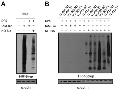
Detection of protein sulfinic acids by NO-Bio in whole cell lysates. (A) Analysis of protein sulfinylation in HeLa cell lysates (5 ug protein loaded per lane). (B) Analysis of protein sulfinylation in human lung tumor tissue lysates (1 ug protein loaded per lane). Legend: T1-001-T1 papillary adenocarcinoma; T1-001-N1 matched normal tissue; T1-005-T1 adenocarcinoma; T1-005-N1 matched normal tissue; T1-013-T1 adeno-squamous cell carcinoma; T1-013-N1 matched normal tissue.
Adapted from: Lo Conte M., et al. ACS Chem. Biol. 2015.
Probe can be used to detect formation of protein sulfinic acid via a two-step approach:
Step 1 - The free cysteine residues are selectively blocked.
Step 2 - The sample is treated with NO-Bio to label sulfinic acids.
NOTE: Stock solution in DMSO (suggested dilution: 25-50 mM) can be added to cell lysis buffer, keeping final [DMSO] <4%.
- Lo Conte M., Lin J., Wilson M.A., Carroll K.S. A Chemical Approach for the Detection of Protein Sulfinylation. ACS Chem. Biol. 2015.
If you publish research with this product, please let us know so we can cite your paper.