Z-Cy2 Maleimide

Thiol mono-reactive zwitterionic and pI balancing Cy2 dye (Z-Cy2 dye). This reagent can be used to attach Cy2 fluorophore to proteins and peptides containing cysteine residues, as well as to other thiolated molecules (such as thiol-containing oligonucleotides).
Highlights:
- Highly water-soluble due to cysteic acid group
- Titratable amine group balances protein pI by compensating for the loss of a cysteine side chain upon labeling
- Enhanced detection sensitivity, compared to traditional CyDyes
- Maleimide functionality
From the laboratories of Edward A. Dratz, PhD, and Paul A. Grieco, PhD, Montana State University.
See more by visiting the Grieco Group Labpage.
 Part of The Investigator's Annexe program.
Thiol mono-reactive zwitterionic and pI balancing Cy2 dye (Z-Cy2 dye). This reagent can be used to attach Cy2 fluorophore to proteins and peptides containing cysteine residues, as well as to other thiolated molecules (such as thiol-containing oligonucleotides).
Highlights:
- Highly water-soluble due to cysteic acid group
- Titratable amine group balances protein pI by compensating for the loss of a cysteine side chain upon labeling
- Enhanced detection sensitivity, compared to traditional CyDyes
- Maleimide functionality
From the laboratories of Edward A. Dratz, PhD, and Paul A. Grieco, PhD, Montana State University.
See more by visiting the Grieco Group Labpage.
![]() Part of The Investigator's Annexe program.
Part of The Investigator's Annexe program.
| Product Type: | Small Molecule |
| Name: | Z-Cy2 Maleimide |
| Chemical Formula: | C44H48N6O11S |
| Molecular Weight: | 869 |
| Format: | Orange powder |
| Purity: | 95+% (NMR, HPLC) |
| Solubility: | Good in water and polar organic solvents |
| Spectral Information: | Excitation maximum: 485nmExtinction at max excitation: 105,000Emission maximum: 500nmFluorescence quantum yield: 0.07 |
| Storage: | -20C. Protect from light. Dessicate. |
| Shipped: | Cold packs |
Application Notes
Suggested amount: 4nmol/50ug protein. NOTE: Cystines should be reduced with TCEP (tris-carboxyethylphosphine) prior to the labeling.
Labeling is performed according to the standard protocol from GE Healthcare. For a protein extract containing 5 µg protein in 9 uL cell lysis buffer (30 mM Tris, 7 M Urea, 2 M Thiourea, 4% (w/v) CHAPS, 5 mM magnesium acetate, pH 8.0), add 1 uL 2 mM TCEP and mix vigorously. Spin down the sample in a microcentrifuge and incubate at 37 °C for 1 hour in the dark. Add 2 uL 2 mM Z-CyDye maleimide and mix vigorously. Spin down the sample in a microcentrifuge and incubate at 37 °C for 30 minutes in the dark. Stop the reaction by adding 12 uL sample buffer (Pharmalytes (2% final), DTT (130 mM final), 7 M Urea, 2 M Thiourea, 4% CHAPS). Mix vigorously by pipetting and spin down the sample in a microcentrifuge.
2D DIGE Examples
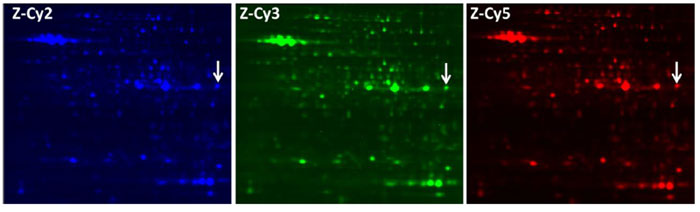
2D gel of total soluble protein from Sulfolobus solfataricus labeled with Z-CyDyes. Each dye was used to label the protein sample separately. Dye labeled protein samples were combined and run on the same gel. After separation, the gel was scanned with 100 ?m steps using the following laser excitation/band-pass filter combinations: 488/520 nm for Z-Cy2 (left), 532/580 nm for Z-Cy3 (middle), and 633/670 nm for Z-Cy5 (right). Images show the relatively crowded central region of the gel (pI 4?9 and 15?150 kDa), revealing well-defined spots with all three dyes.
Adapted from: Epstein MG, et al. Bioconjug Chem. 2013 Sep 18;24(9):1552-61.
Comparison of Total Spot Number
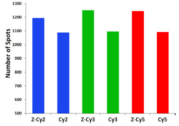
Total number of spots detected from a gel using Z-Cy2, ZCy3, and Z-Cy5 and from a matched gel using CyDye DIGE Fluor minimal Cy2, Cy3, and Cy5 purchased from GE Healthcare. The protein samples labeled and experimental protocols were the same for both gels and all dyes. Progenesis SameSpots software detects more spots when Z-CyDyes are used in comparison to CyDyes.
Adapted from: Epstein MG, et al. Bioconjug Chem. 2013 Sep 18;24(9):1552-61.
Looking for a different Z-Cy Dye? Check out our other Z-Cy Dyes!
- Epstein MG, Reeves BD, Maaty WS, Fouchard D, Dratz EA, Bothner B, Grieco PA. Enhanced sensitivity employing zwitterionic and pI balancing dyes (Z-CyDyes) optimized for 2D-gel electrophoresis based on side chain modifications of CyDye fluorophores. New tools for use in proteomics and diagnostics. Bioconjug Chem. 2013 Sep 18;24(9):1552-61.
- Reeves BD, Joshi N, Campanello GC, Hilmer JK, Chetia L, Vance JA, Reinschmidt JN, Miller CG, Giedroc DP, Dratz EA, Singel DJ, Grieco PA. Conversion of S-phenylsulfonylcysteine residues to mixed disulfides at pH 4.0: utility in protein thiol blocking and in protein-S-nitrosothiol detection. Org Biomol Chem. 2014 Jul 2.
If you publish research with this product, please let us know so we can cite your paper.