Goat Anti-PPP1R12A Antibody

This goat IgG polyclonal antibody was generated against peptide sequence CREDEYKQKYSRTYD from the C Terminus of protein phosphatase 1 regulatory subunit 12A (PPP1R12A) and recognizes human PPP1R12A.
Highlights:
- Reacts with human PPP1R12A
- Suitable for Peptide ELISA, Immunohistochemistry, and Immunofluorescence applications
- Associated with Genitourinary And/Or Brain Malformation Syndrome
Protein phosphatase 1 regulatory subunit 12A (PPP1R12A) interacts with protein phosphatase type 1 catalytic unit (PP1C) and M20/21 to form the trimeric holoenzyme, myosin phosphatase (MP). Myosin phosphatase is a key regulator of cell morphology and motility. Phosphorylation of specific sites on PPP1R12A inhibits its activity with pathogenic variants of the PPP1R12A gene resulitng in a non-functional MP. Diseases associated with PPP1R12A include Genitourinary And/Or Brain Malformation Syndrome.
| Product Type: | Antibody |
| Name: | Goat Anti-PPP1R12A Antibody |
| Alternative Name(s): | Myosin phosphatase-targeting subunit 1 (Myosin phosphatase target subunit 1), Protein phosphatase myosin-binding subunit, MBS, MYPT1 |
| Accession ID: | NP_002471.1, NP_001231919.1, NP_001231921 .1, NP_001137358.1 |
| Antigen: | PPP1R12A |
| Isotype: | IgG |
| Clonality: | Polyclonal |
| Reactivity: | Human |
| Specificity: | PPP1R12A |
| Immunogen: | CREDEYKQKYSRTYD |
| Species Immunized: | Goat |
| Epitope: | C Terminus |
| Purification Method: | Purified from goat serum by ammonium sulphate precipitation followed by antigen affinity chromatography using the immunizing peptide |
| Buffer: | Supplied at 0.5 mg/ml in Tris saline, 0.02% sodium azide, pH7.3 with 0.5% bovine serum albumin. |
| Tested Applications: | Pep-ELISA, IHC, IF |
| Storage: | Aliquot and store at -20C. Minimize freezing and thawing. |
| Shipped: | Cold Packs |
Immunofluorescence and Immunohistochemistry
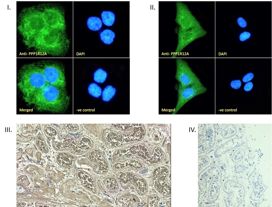
I. Immunofluorescence analysis of paraformaldehyde fixed A431 cells, permeabilized with 0.15% Triton. Primary incubation 1hr (10ug/ml) followed by Alexa Fluor 488 secondary antibody (2ug/ml), showing cytoplasmic staining. The nuclear stain is DAPI (blue). Negative control: Unimmunized goat IgG (10ug/ml) followed by Alexa Fluor 488 secondary antibody (2ug/ml). II. Immunofluorescence analysis of paraformaldehyde fixed U2OS cells, permeabilized with 0.15% Triton. Primary incubation 1hr (10ug/ml) followed by Alexa Fluor 488 secondary antibody (2ug/ml), showing cytoplasmic staining. The nuclear stain is DAPI (blue). Negative control: Unimmunized goat IgG (10ug/ml) followed by Alexa Fluor 488 secondary antibody (2ug/ml). III. (4µg/ml) staining of paraffin embedded Human Testis. Heat induced antigen retrieval with citrate buffer pH 6, HRP-staining. IV. Negative control showing staining of paraffin embedded Human Testis with no primary antibody.
If you publish research with this product, please let us know so we can cite your paper.