Goat Anti-EEA1 (aa821-835) Antibody
 Antibody")
This goat IgG polyclonal antibody was generated against peptide sequence CQETKIQHEELNNRIQ from the Internal Region of early endosome antigen 1 (EEA1) and recognizes human and mouse EEA1.
Highlights:
- Reacts with human and mouse EEA1
- Suitable for Peptide ELISA, Western Blot, and Immunofluorescence applications
- Associated with Subacute Cutaneous Lupus Erythematosus and Cutaneous Lupus Erythematosus
Early endosomes are involved in receiving endocytosed materials and sorting them for transport to late endosomes or recycling them to the plasma membrane. Early endosome antigen 1 (EEA1) is associated with early endosomes and is involved in endosome trafficking by binding phospholipid vesicles containing phosphatidylinositol 3-phosphate. Diseases associated with EEA1 include Subacute Cutaneous Lupus Erythematosus and Cutaneous Lupus Erythematosus.
| Product Type: | Antibody |
| Name: | Goat Anti-EEA1 (aa821-835) Antibody |
| Alternative Name(s): | Endosome-associated protein p162, Zinc finger FYVE domain-containing protein 2, ZFYVE2 |
| Accession ID: | NP_003557.2 |
| Antigen: | EEA1 (aa821-835) |
| Isotype: | IgG |
| Clonality: | Polyclonal |
| Reactivity: | Human, Mouse |
| Specificity: | EEA1 (aa821-835) |
| Immunogen: | CQETKIQHEELNNRIQ |
| Species Immunized: | Goat |
| Epitope: | Internal Region |
| Purification Method: | Purified from goat serum by ammonium sulphate precipitation followed by antigen affinity chromatography using the immunizing peptide |
| Buffer: | Supplied at 0.5 mg/ml in Tris saline, 0.02% sodium azide, pH7.3 with 0.5% bovine serum albumin. |
| Tested Applications: | Pep-ELISA, WB, IF |
| Storage: | Aliquot and store at -20C. Minimize freezing and thawing. |
| Shipped: | Cold Packs |
Western Blot
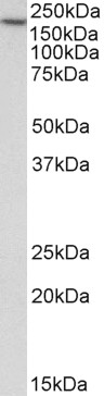
(0.3µg/ml) staining of HEK293 lysate (35µg protein in RIPA buffer). Detected by chemiluminescence.
Immunofluorescence
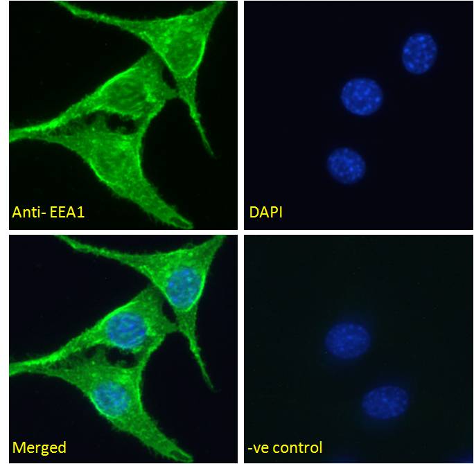
Immunofluorescence analysis of paraformaldehyde fixed NIH3T3 cells, permeabilized with 0.15% Triton. Primary incubation 1hr (5ug/ml) followed by Alexa Fluor 488 secondary antibody (2ug/ml), showing vesicle/cytoplasmic staining. The nuclear stain is DAPI (blue). Negative control: Unimmunized goat IgG (5ug/ml) followed by Alexa Fluor 488 secondary antibody (2ug/ml).
If you publish research with this product, please let us know so we can cite your paper.