Goat Anti-GAPDH (C Terminus) Loading Control Antibody
 Loading Control Antibody")
This goat IgG polyclonal antibody was generated against peptide sequence CHQVVSSDFNSDT from the C terminus of glyceraldehyde-3-phosphate dehydrogenase (GAPDH) and recognizes human and rat GAPDH.
Highlights:
- Reacts with human and rat GAPDH
- Suitable for Peptide ELISA, Western Blot, Immunofluorescence, and Immunohistochemistry applications
- Useful as a loading or positive control
GAPDH is constitutively expressed in almost all tissues at high levels. It is therefore a useful marker when a loading or positive control is required in an assay.
| Product Type: | Antibody |
| Name: | Goat Anti-GAPDH (C Terminus) Loading Control Antibody |
| Alternative Name(s): | Peptidyl-cysteine S-nitrosylase GAPDH, GAPD |
| Accession ID: | NP_002037.2 |
| Antigen: | GAPDH (C Terminus) Loading Control |
| Isotype: | IgG |
| Clonality: | Polyclonal |
| Reactivity: | Human, Rat |
| Specificity: | GAPDH (C Terminus) Loading Control |
| Immunogen: | CHQVVSSDFNSDT |
| Species Immunized: | Goat |
| Epitope: | C Terminus |
| Purification Method: | Purified from goat serum by ammonium sulphate precipitation followed by antigen affinity chromatography using the immunizing peptide |
| Buffer: | Supplied at 0.5 mg/ml in Tris saline, 0.02% sodium azide, pH7.3 with 0.5% bovine serum albumin. |
| Tested Applications: | Pep-ELISA, WB, IF, IHC |
| Storage: | Aliquot and store at -20C. Minimize freezing and thawing. |
| Shipped: | Cold Packs |
Western Blot, Immunofluorescence, and Immunohistochemistry
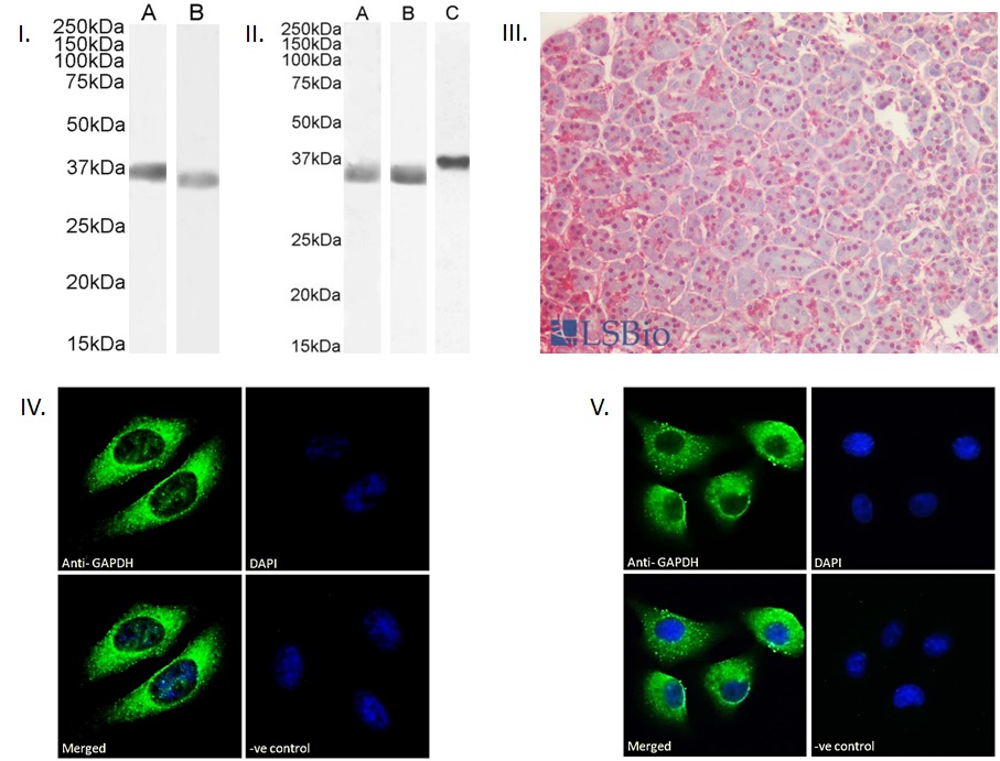
I. (0.001µg/ml) staining of HEK293 (A) and HeLa (B) cell lysate (35µg protein in RIPA buffer). Detected by chemiluminescence. II. (0.001µg/ml) staining of Human Liver (A), Tonsil (B) and (0.3ug/ml) Rat Brain (C) lysate (35µg protein in RIPA buffer). Detected by chemiluminescence. III. (2.5µg/ml) staining of paraffin embedded Human Pancreas. Steamed antigen retrieval with citrate buffer pH 6, AP-staining. IV. Immunofluorescence analysis of paraformaldehyde fixed A549 cells, permeabilized with 0.15% Triton. Primary incubation 1hr (10ug/ml) followed by Alexa Fluor 488 secondary antibody (2ug/ml), showing cytoplasmic and vesicle staining. The nuclear stain is DAPI (blue). Negative control: Unimmunized goat IgG (10ug/ml) followed by Alexa Fluor 488 secondary antibody (2ug/ml). V. Immunofluorescence analysis of paraformaldehyde fixed HeLa cells, permeabilized with 0.15% Triton. Primary incubation 1hr (10ug/ml) followed by Alexa Fluor 488 secondary antibody (2ug/ml), showing cytoplasmic and vesicle staining. The nuclear stain is DAPI (blue). Negative control: Unimmunized goat IgG (10ug/ml) followed by Alexa Fluor 488 secondary antibody (2ug/ml).
- Kiepe D, Van Der Pas A, Ciarmatori S, Ständker L, Schütt B, Hoeflich A, Hügel U, Oh J, Tönshoff B., Defined carboxy-terminal fragments of insulin-like growth factor (IGF) binding protein 2 exert similar mitogenic activity on cultured rat growth plate chondrocytes as IGF-I., Endocrinology. 2008 Oct;149(10):4901-11. ,18556354
If you publish research with this product, please let us know so we can cite your paper.