Goat Anti-VPS35 / MEM3 Antibody

This goat IgG polyclonal antibody was generated against peptide sequence CSPESEGPIYEGLIL from the C Terminus of vacuolar protein sorting-associated protein 35 (VPS35) and recognizes human, mouse, and rat VPS35.
Highlights:
- Reacts with human, mouse, and rat VPS35
- Suitable for Peptide ELISA, Western Blot, Immunohistochemistry, and Immunofluorescence applications
- Parkinson disease 17 and hereditary late-onset Parkinson disease are associated with mutations in the VPS35 gene.
Vacuolar protein sorting-associated protein 35 (VPS35) is a component of a multimeric complex termed the retromer cargo-selective complex (CSC). In addition to VPS35, the CSC is comprised of two other proteins, VPS26 and Vps29. The CSC is involved in the retrograde transport of cargo proteins from endosomes to the trans-Golgi network predominantly via VPS35. Mutations in the VPS35 gene are associated with Parkinson disease 17 and hereditary late-onset Parkinson disease.
| Product Type: | Antibody |
| Name: | Goat Anti-VPS35 / MEM3 Antibody |
| Alternative Name(s): | MEM3, PARK17 |
| Accession ID: | NP_060676.2 |
| Antigen: | VPS35 / MEM3 |
| Isotype: | IgG |
| Clonality: | Polyclonal |
| Reactivity: | Human, Mouse, Rat |
| Specificity: | VPS35 / MEM3 |
| Immunogen: | CSPESEGPIYEGLIL |
| Species Immunized: | Goat |
| Epitope: | C Terminus |
| Purification Method: | Purified from goat serum by ammonium sulphate precipitation followed by antigen affinity chromatography using the immunizing peptide |
| Buffer: | Supplied at 0.5 mg/ml in Tris saline, 0.02% sodium azide, pH7.3 with 0.5% bovine serum albumin. |
| Tested Applications: | Pep-ELISA, WB, IHC, IF |
| Storage: | Aliquot and store at -20C. Minimize freezing and thawing. |
| Shipped: | Cold Packs |
Western Blot, Immunofluorescence, and Immunohistochemistry
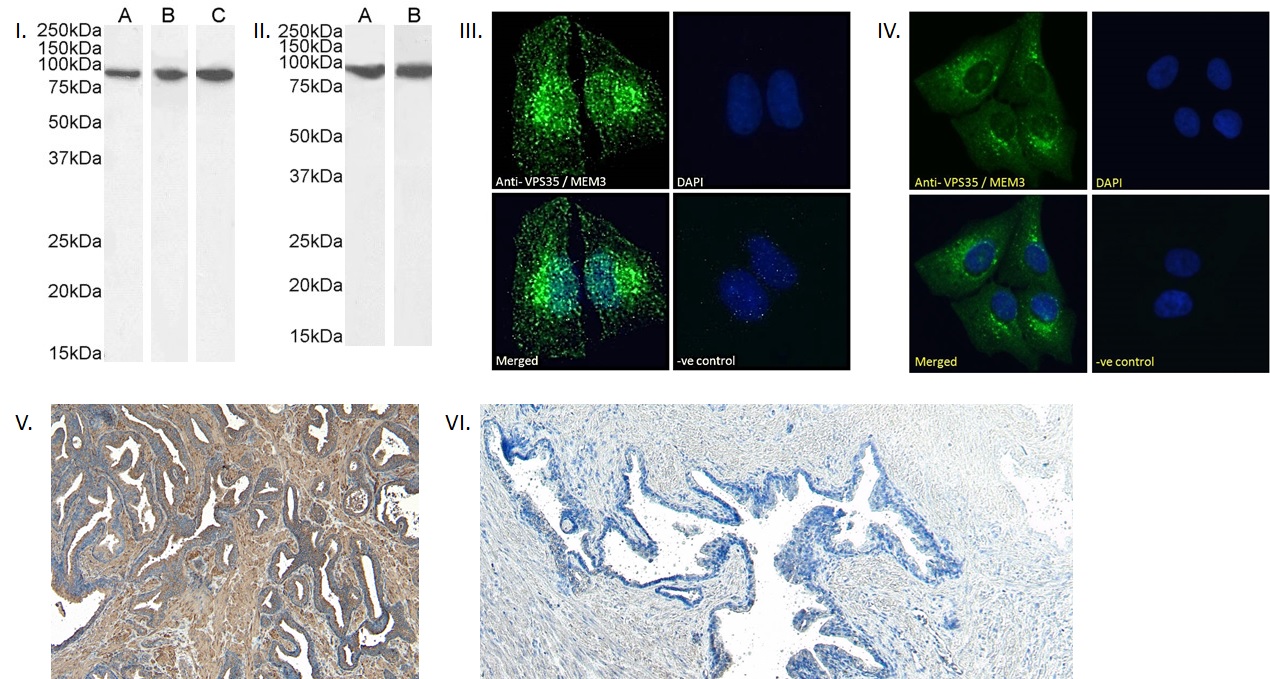
I. (0.03µg/ml) staining of Human (A) Mouse (B) and Rat (C) Brain lysate (35µg protein in RIPA buffer). Detected by chemiluminescence. II. (0.03µg/ml) staining of HepG2 (A) and HEK293 (B) cell lysate (35µg protein in RIPA buffer). Detected by chemiluminescence. III. Immunofluorescence analysis of paraformaldehyde fixed HEK293 cells, permeabilized with 0.15% Triton. Primary incubation 1hr (10ug/ml) followed by Alexa Fluor 488 secondary antibody (2ug/ml), showing cytoplasmic/vesicle staining. The nuclear stain is DAPI (blue). Negative control: Unimmunized goat IgG (10ug/ml) followed by Alexa Fluor 488 secondary antibody (2ug/ml). IV. Immunofluorescence analysis of paraformaldehyde fixed U2OS cells, permeabilized with 0.15% Triton. Primary incubation 1hr (10ug/ml) followed by Alexa Fluor 488 secondary antibody (2ug/ml), showing cytoplasmic/vesicle staining. The nuclear stain is DAPI (blue). Negative control: Unimmunized goat IgG (10ug/ml) followed by Alexa Fluor 488 secondary antibody (2ug/ml). V. (8µg/ml) staining of paraffin embedded Human Prostate. Heat induced antigen retrieval with citrate buffer pH 6, HRP-staining. VI. Negative control showing staining of paraffin embedded Human Prostate with no primary antibody.
- Lee S, Uchida Y, Emoto K, Umeda M, Kuge O, Taguchi T, Arai H., Impaired retrograde membrane traffic through endosomes in a mutant CHO cell defective in phosphatidylserine synthesis., Genes Cells. 2012 Aug;17(8):728-36. doi: 10.1111/j.1365-2443.2012.01622.x. ,22747682
If you publish research with this product, please let us know so we can cite your paper.