Goat Anti-Smooth Muscle Alpha-Actin Antibody

This goat IgG polyclonal antibody was generated against peptide sequence EEEDSTALVC from the N Terminus of smooth muscle alpha-actin (ACTA2) and recognizes human and mouse ACTA2.
Highlights:
- Reacts with human and mouse ACTA2
- Suitable for Peptide ELISA and Western Blot applications
- Mutations are associated with thoracic aortic disease, premature coronary artery disease, ischaemic strokes, Moyamoya disease, and multisystemic smooth muscle dysfunction syndrome
Actins are a family of six ubiquitously expressed proteins involved in a number of cellular functions including muscle contraction, cell motility, maintenance of cell shape, and regulation of transcription. In vertebrates, three actin isoforms, alpha, beta, and gamma, have been identified. Smooth muscle alpha-actin (ACTA2) is a smooth muscle actin involved in vascular contractility and blood pressure homeostasis. Mutations in this gene are associated with a variety of diseases such as thoracic aortic disease, premature coronary artery disease, ischaemic strokes, Moyamoya disease, and multisystemic smooth muscle dysfunction syndrome.
| Product Type: | Antibody |
| Name: | Goat Anti-Smooth Muscle Alpha-Actin Antibody |
| Alternative Name(s): | ACTSA, ACTVS |
| Accession ID: | NP_001604.1; NP_001135417.1 |
| Antigen: | Smooth muscle alpha-actin |
| Isotype: | IgG |
| Clonality: | Polyclonal |
| Reactivity: | Human, Mouse |
| Specificity: | Smooth muscle alpha-actin |
| Immunogen: | EEEDSTALVC |
| Species Immunized: | Goat |
| Epitope: | N Terminus |
| Purification Method: | Purified from goat serum by ammonium sulphate precipitation followed by antigen affinity chromatography using the immunizing peptide |
| Buffer: | Supplied at 0.5 mg/ml in Tris saline, 0.02% sodium azide, pH7.3 with 0.5% bovine serum albumin. |
| Tested Applications: | Pep-ELISA, WB |
| Storage: | Aliquot and store at -20C. Minimize freezing and thawing. |
| Shipped: | Cold Packs |
Western Blot
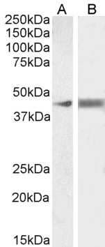
(0.5µg/ml) staining of Human (A) and Rat (B) Duodenum lysate (35µg protein in RIPA buffer). Detected by chemiluminescence.
Western Blot
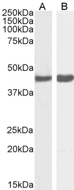
(0.5µg/ml) staining of HeLa (A) and (1ug/ml) NIH3T3 (B) cell lysate (RIPA buffer, (35µg protein in RIPA buffer). Detected by chemiluminescence.
- McIntire RH, Sifers T, Platt JS, Ganacias KG, Langat DK, Hunt JS., Novel HLA-G-binding leukocyte immunoglobulin-like receptor (LILR) expression patterns in human placentas and umbilical cords., Placenta. 2008 Jul;29(7):631-8. ,18538388
If you publish research with this product, please let us know so we can cite your paper.