Goat Anti-AIF1 / IBA1 (Isoform 1 and 3) Antibody
 Antibody")
This goat IgG polyclonal antibody was generated against peptide sequence C-TGPPAKKAISELP from the C Terminus of allograft inflammatory factor 1 (AIF1 / IBA1) and recognizes human, mouse, and rat AIF1 / IBA1 (isoform 1 and 3).
Highlights:
- Reacts with human, mouse, and rat AIF1 / IBA1
- This antibody is expected to recognize isoform 1 (NP_116573.1) and isoform 3 (NP_001614.3)
- Suitable for Peptide ELISA, Western Blot, and Immunohistochemistry applications
Allograft inflammatory factor 1 (AIF1), also known as ionized calcium-binding adapter molecule 1 (IBA1), is a cytosolic protein that binds actin and calcium. AIF1 expression is induced by cytokines and interferon. Expression of this protein is upregulated in activated macrophages and may play a role in cancer progression, rheumatoid arthritis, renal disease, and retinal disease.
| Product Type: | Antibody |
| Name: | Goat Anti-AIF1 / IBA1 (Isoform 1 and 3) Antibody |
| Alternative Name(s): | IBA1, IRT1, AIF-1, IRT-1 |
| Accession ID: | NP_116573.1; NP_001614.3 |
| Antigen: | AIF1 / IBA1 (Isoform 1 and 3) |
| Isotype: | IgG |
| Clonality: | Polyclonal |
| Reactivity: | Human, Mouse, Rat |
| Specificity: | AIF1 / IBA1 (Isoform 1 and 3) |
| Immunogen: | CTGPPAKKAISELP |
| Species Immunized: | Goat |
| Epitope: | C Terminus |
| Purification Method: | Purified from goat serum by ammonium sulphate precipitation followed by antigen affinity chromatography using the immunizing peptide |
| Buffer: | Supplied at 0.5 mg/ml in Tris saline, 0.02% sodium azide, pH7.3 with 0.5% bovine serum albumin. |
| Tested Applications: | Pep-ELISA, WB, IHC |
| Storage: | Aliquot and store at -20C. Minimize freezing and thawing. |
| Shipped: | Cold Packs |
Western Blot and Immunohistochemistry
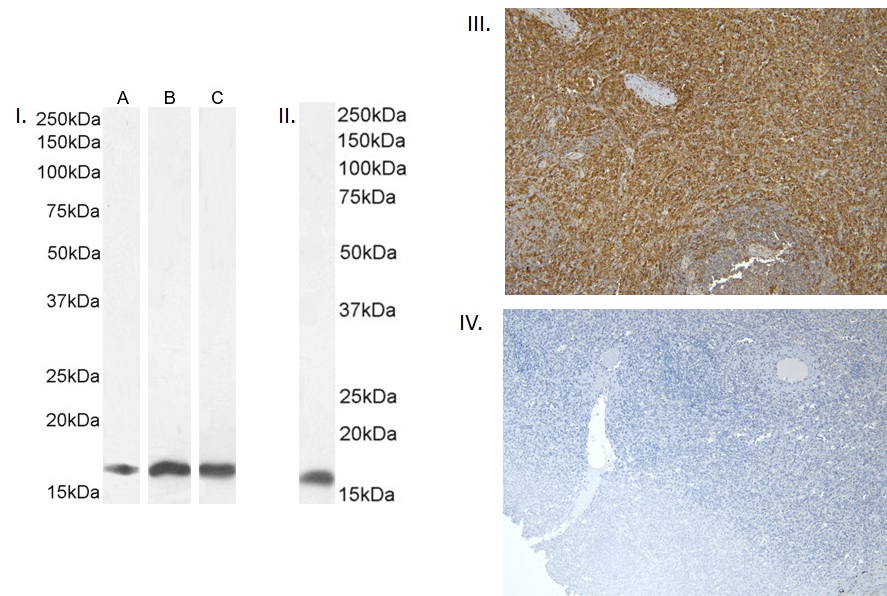
I. (1µg/ml) staining of Human Frontal Cortex (A) and (0.1ug/ml) Mouse (B) and Rat (C) Brain lysate (35µg protein in RIPA buffer). Detected by chemiluminescence. II. (0.1µg/ml) staining of Mouse Lymph node lysate (35µg protein in RIPA buffer). Detected by chemiluminescence. III. (8µg/ml) staining of paraffin embedded Human Spleen. Heat induced antigen retrieval with citrate buffer Ph 6, HRP-staining. IV. Negative control showing staining of paraffin embedded Human Spleen with no primary antibody.
- Recio JS, Álvarez-Dolado M, Díaz D, Baltanás FC, Piquer-Gil M, Alonso JR, Weruaga E., Bone marrow contributes simultaneously to different neural types in the central nervous system through different mechanisms of plasticity., Cell Transplant. 2011;20(8):1179-92. ,21294954
If you publish research with this product, please let us know so we can cite your paper.