Anti-Lamin B Receptor (LBR) [BB2SS3F3] Antibody
![Anti-Lamin B Receptor (LBR) [BB2SS3F3] Antibody, 5mL (supernatant)](https://www.kerafast.com/MediaStorage/Product/Images/Large/1744_2001202001265917430.jpg "Anti-Lamin B Receptor (LBR) [BB2SS3F3] Antibody")
This mouse IgG1, kappa monoclonal antibody was raised against glutathione-S-transferase fused to C-terminal fragment of mouse lamin B2 (amino acids residues 380-596) and recognizes human and mouse Lamin B Receptor (LBR).
Highlights:
- Reacts with human and mouse Lamin B Receptor (LBR)
- Suitable for Western Blot, Immunohistochemistry, Immunocytochemistry and Immunofluorscence applications
Lamins are intermediate filament proteins found inside the nuclei of metazoans. They form a filamentous meshwork concentrated at the nuclear envelope periphery and play an important role in nuclear structure and many DNA-related processes. Two classes of lamins are distinguished: A-type (lamins A and C) and B-type (lamins B1 and B2). The ubiquitously expressed lamin B2 is a product of LMNB2 gene and is essential for survival in mice. Mice deficient in lamin B2 die soon after birth with brain abnormalities caused by defects in neuronal migration. In humans, mutations in LMNB2 gene have been associated with progressive myoclonus epilepsy and partial lipodystrophy. Of note, a shorter spliced variant of LMNB2 gene is expressed in testis and is known as lamin B3.
From the laboratory of Brian Burke, PhD, A*STAR Institute of Medical Biology (IMB).
| Product Type: | Antibody |
| Accession ID: | Q14739 |
| Antigen: | LBR |
| Molecular Weight: | Human LBR 70.7kDa; mouse LBR 71.4kDa |
| Isotype: | IgG1 kappa |
| Clonality: | Monoclonal |
| Clone Name: | BB2SS3F3 |
| Reactivity: | Human and Mouse |
| Immunogen: | Glutathione-S-transferase fused to C-terminal fragment of mouse lamin B2 (amino acids residues 380-596) |
| Species Immunized: | Mouse |
| Buffer: | Cell culture supernatant |
| Positive Control: | Hela, C2C12, NIE-115 |
| Tested Applications: | WB, IF, IHC, ICC |
| Comments: | Knockout validatedo |
| Storage: | -80C |
| Shipped: | Cold Packs |
Western blot analysis of BB2SS3F3 antibody.
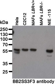
Whole cell lysates from human (Hela) and mouse (C2C12, immortalized mouse adult fibroblasts (MAFs (7)), NIE-115) cell lines were separated on SDS-PAGE gel, transferred to nitrocellulose membrane and blotted with BB2SS3F3 antibody. Both primary and secondary antibodies were diluted in blocking buffer (5% nonfat dry milk resuspended in 0.1% Tween and 1X PBS). The predicted molecular weight is 70.7kDA and 71.4kDa for human and mouse LBR respectively. The absence of a band in LBR-null MAFs indicates that the antibody specifically recognizes LBR.
Immunofluorescence staining of small intestine from adult C57BL/6 mice.
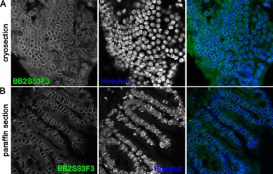
(A) Small intestine was transferred to OCT compound and frozen in liquid nitrogen. Tissue cryosections were fixed in 10% neutral buffered formalin, were incubated with 1% Triton X-100 (in PBS) and stained with anti-LBR BB2SS3F3 antibody. (B) Tissue was fixed in 10% NBF and then embedded in paraffin. Following heat-induced antigen retrieval (Sodium citrate buffer pH 6) the paraffin sections were incubated with 1% Triton X-100 (in PBS) and subsequently stained with mouse monoclonal anti-LBR BB2SS3F3 antibody. Nuclei were visualized with Hoechst.
Confocal images of human (Hela) and mouse (NIE-115) cell lines immunofluorescently stained with BB2SS3F3 antibody.
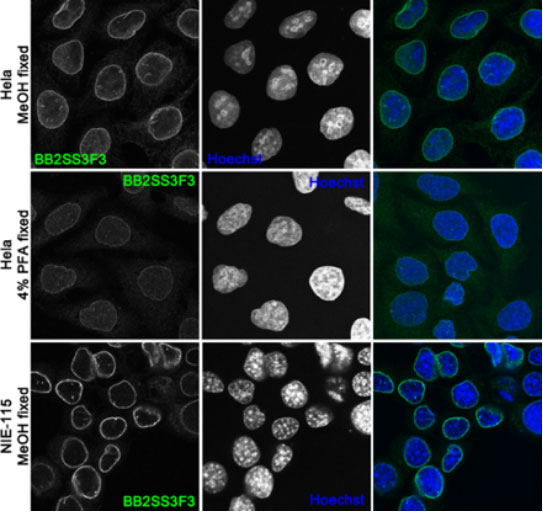
The BB2SS3F3 antibody works on cells fixed with 4% PFA and cold methanol with methanol fixation resulting in brighter signal of the antibody. Nuclei were counterstained with Hoechst.
The antibody specifically detects LBR.
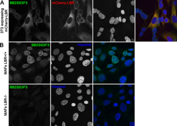
(A) 3T3 cells stably expressing mCherry-tagged mouse LBR were fixed in 4% PFA and stained with BB2SS3F3 antibody. The expression levels of mCherry-LBR correspond to intensity of BB2SS3F3 antibody staining. (B) Immortalized wild-type mouse adult fibroblast (MAFs LBR+/+) (upper panel) and LBR-null MAFs (MAFs LBR-/-) (7) (lower panel) were methanol fixed and immunofluorescently stained with BB2SS3F3 mouse monoclonal antibody. In LBR-null MAFs there is a loss of BB2SS3F3 antibody staining signal indicating antibody specificity for LBR.
From the laboratory of Brian Burke, PhD, A*STAR Institute of Medical Biology (IMB).
Immunocytochemistry/Immunofluorescence: formaldehyde, methanol fixation with methanol fixation resulting in a much brighter signal
- Burke, B., C.L. Stewart. 2013. The nuclear lamins: flexibility in function. Nat Rev Mol Cell Biol. 14(1):13-24.
- Coffinier, C., S.Y. Chang, C. Nobumori, Y. Tu, E.A. Farber, J.I. Toth, L.G. Fong, S.G. Young. 2010. Abnormal development of the cerebral cortex and cerebellum in the setting of lamin B2 deficiency. Proc Natl Acad Sci USA. 107:5076-5081.
- Kim, Y., A.A. Sharov, K. McDole, M. Cheng, H. Hao, C.M. Fan, N. Gaiano, M.S. Ko, Y. Zheng. 2011. Mouse B-type lamins are required for proper organogenesis but not by embryonic stem cells. Science. 334(6063):1706-10.
- Hegele, R. A., H. Cao, D. M. Liu, G. A Costain, V. Charlton-Menys, N. W. Rodger, P.N Durrington. 2006. Sequencing of the reannotated LMNB2 gene reveals novel mutations in patients with acquired partial lipodystrophy. Am J Hum Genet. 79(2):383-9 5.
- Damiano, J.A., Z. Afawi, M. Bahlo, M. Mauermann, A. Misk, T. Arsov, K.L. Oliver, H.H. Dahl, A.E. Shearer, R.J. Smith et al. 2015. Mutation of the nuclear lamin gene LMNB2 in progressive myoclonus epilepsy with early ataxia. Hum Mol Genet. 24:4483-4490.
If you publish research with this product, please let us know so we can cite your paper.