Anti-Lamin A+C [LASS2D9] Antibody
![Anti-Lamin A+C [LASS2D9] Antibody, 5mL (supernatant)](https://www.kerafast.com/MediaStorage/Product/Images/Large/1743_2001202001265917420.jpg "Anti-Lamin A+C [LASS2D9] Antibody")
This mouse IgG3 kappa monoclonal antibody recognizes human and mouse Lamin A+C.
Highlights:
- Reacts with human and mouse lamin A+C
- Suitable for Western Blot, Immunocytochemistry, Immunohistochemistry and Immunofluorscence applications
Lamins A and C are major components of the nuclear lamina, a thin protein meshwork located at the nuclear face of the nuclear envelope (NE). The lamina provides structural integrity to the NE and is involved in many other aspects of nuclear biology including transcription and chromatin organization. Lamins A and C arise from the LMNA gene by alternative splicing and majority of adult tissues express at least one of the isoform. The LMNA gene has been extensively studies due to its association with variety of human diseases. Mutations in LMNA have been linked 12 distinct disorders, including Emery-Dreifus muscular dystrophy, dilated cardiomyopathy, Dunnigan-type partial lipodystrophy and Hutchinson-Gilford progeria syndrome.
From the laboratory of Brian Burke, PhD, A*STAR Institute of Medical Biology (IMB).
| Product Type: | Antibody |
| Antigen: | Lamin A+C |
| Accession ID: | PO2545 |
| Molecular Weight: | Lamin A 74kDa, Lamin C 65 kDa |
| Isotype: | IgG3 kappa |
| Clonality: | Monoclonal |
| Clone Name: | LASS2D9 |
| Reactivity: | Human, mouse, not tested with other species |
| Immunogen: | Unrelated antigen |
| Species Immunized: | Mouse |
| Buffer: | Cell culture supernatant |
| Tested Applications: | WB, IF, IHC, ICC |
| Positive Control: | Hela, 293T, C2C12, fibroblasts |
| Storage: | -80C |
| Shipped: | Cold Packs |
Western Blot
Western blot analysis of LASS2D9 antibody. Whole cell lysate from human (Hela) and mouse (C2C12, mouse adult fibroblasts (MAFs)) cell lines cells were separated on SDS-PAGE gel, transferred to nitrocellulose membrane and blotted with LASS2D9 antibody. The membrane was blocked overnight in blocking buffer (5% nonfat dry milk resuspended in 0.1% Tween and 1X PBS). Both primary and secondary antibodies were diluted in blocking buffer. The predicted molecular weight for lamin A is 74 kDA and for lamin C 65 kDa. The absence of a band in LMNA null MAFs indicated that the antibody specifically recognizes products of LMNA gene.
Immunofluorescence staining of mouse small intestine.
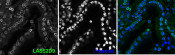
Small intestine from adult C57BL/6 mice was transferred to OCT compound and frozen in liquid nitrogen. Tissue cryosections were fixed in 10% neutral buffered formalin, permeabilized and stained with anti-lamin A+C LASS2D9 antibody.
Immunofluorescence staining of human (Hela) and mouse (C2C12) cell lines with LASS2D9 antibody.
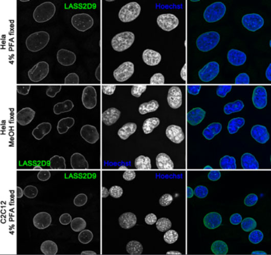
Cells were fixed in 4% PFA (upper and bottom panels) or cold methanol (middle panel) and labelled with mouse monoclonal LASS2D9 antibody.
The mouse monoclonal LASS2D9 antibody specifically detects lamin A and lamin C.
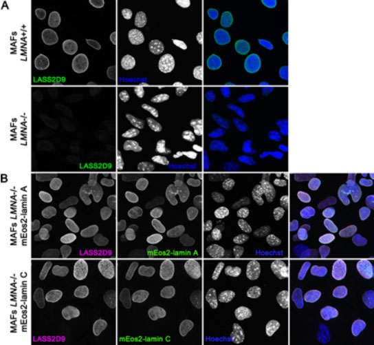
(A) Wild-type mouse adult fibroblast (MAFs LMNA+/+) (upper panel) and LMNA-null MEFs (MAFs LMNA-/-) (lower panel) were methanol fixed and immunofluorescently stained with LASS2D9 mouse monoclonal antibody. (B) LMNA-null mouse adult fibroblast stably expressing mEos2-lamin A (upper panel) or mEos2-lamin C (2) (lower panel) were fixed in cold methanol and stained with LASS2D9 antibody (magenta). Expression of the respective LMNA isoform is shown in green. The nuclei were counter-stained with Hoechst. In LMNA-null MEFs there is a loss of LASS2D9 antibody staining signal. Rescue of LMNA-null cells with lamin A or lamin C expressing constructs restores the LASS2D9 antibody signal strongly suggesting that LASS2D9 specifically detects the products of LMNA gene.
From the laboratory of Brian Burke, PhD, A*STAR Institute of Medical Biology (IMB).
- Burke, B., C.L Stewart. 2013. The nuclear lamins: flexibility in function. Nat Rev Mol Cell Biol. 14(1):13-24.
- Xie, W., A. Chojnowski, T. Boudier, J. S. Lim, S. Ahmed, Z. Ser, C. Stewart, B. Burke. 2016. Atype Lamins Form Distinct Filamentous Networks with Differential Nuclear Pore Complex Associations. Curr Biol. 2016 Sep 9. pii: S0960-9822(16)30842-9. doi: 10.1016/j.cub.2016.07.049.
If you publish research with this product, please let us know so we can cite your paper.