Anti-MERS Nucleocapsid (N) [9H10.2.3] Antibody
![Anti-MERS Nucleocapsid (N) [9H10.2.3] Antibody](https://www.kerafast.com/MediaStorage/Product/Images/Large/1705_2001202001265817040.jpg "Anti-MERS Nucleocapsid (N) [9H10.2.3] Antibody")
This mouse monoclonal antibody was raised against recombinant GST-Middle East respiratory syndrome corovirus (MERS-CoV) N fusion protein and recognizes MERS-CoV nucleocapsid protein (MERS-N).
Highlights:
- Reacts with MERS-CoV nucleocapsid protein (MERS-N)
- Suitable for Western Blot, Immunofluorescence and Immunoprecipitation applications
A novel coronavirus (termed as severe acute respiratory syndrome coronavirus, SARS-CoV) was the cause of a viral outbreak which caused profound disturbances worldwide in 2003. Most recently, another novel coronavirus has achieved successful zoonotic transmission and been named as the Middle East respiratory syndrome coronavirus (MERS-CoV). As of 9 May 2014, a total of 536 people have been infected and the fatality rate is ~27%. As the Nucleocapsid protein (N) protein of coronavirus is an abundant virion protein and highly immunogenic, monoclonal antibody for MERS N will be useful for the detection of the virus especially if the monoclonal antibody can distinguish MERS-CoV from other human coronaviruses.
From the laboratory of A*STAR Institute of Molecular and Cell Biology (IMCB) Monoclonal Antibody Unit.
| Product Type: | Antibody |
| Antigen: | MERS-CoV nucleocapsid protein (MERS-N) |
| Accession ID: | K9N4V7 |
| Molecular Weight: | 50 kDa |
| Clonality: | Monoclonal |
| Clone Name: | 9H10.2.3 |
| Reactivity: | MERS-CoV |
| Immunogen: | Recombinant GST-MERS N fusion protein |
| Species Immunized: | Mouse |
| Buffer: | Cell culture supernatant |
| Tested Applications: | WB, IP |
| Storage: | -80C |
| Shipped: | Cold Packs |
Immunofluorescence
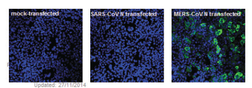
Cos7 cells were mock-transfected (left panel), transfected with plasmid containing either the SARS N (middle panel) or MERS N (right panel) open reading frame. At 24-48h post transfection, cells were fixed with 4% paraformaldehyde, permeabilized with 0.1% Triton X-100 and stained using MERS N monoclonal antibody and followed by secondary goat antimouse FITC (1:100). Nucleic acid staining was done by using DAPI (1ug/ml).
Western Blot
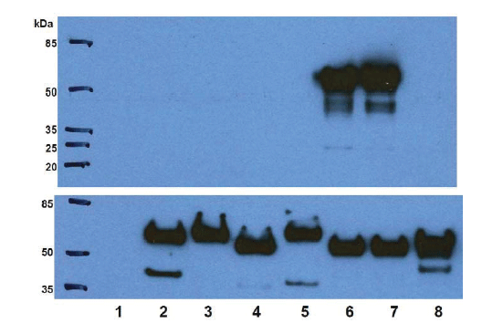
Genes encoding full length N proteins (with a N-terminal myc-tag) of different human coronaviruses were over-expressed in 293FT cells, followed by lysis of cells in RIPA buffer,Western blotting was performed using MERS N monoclonal antibody (upper panel) or mycmonoclonal antibody (lower panel). As shown, the MERS N monoclonal antibody bound specifically to the N protein from MERS-CoV (lanes 6 and 7) but not to N proteins of other coronavirus. Lane 1: Untransfected, Lane 2: 229E-N, Lane 3: HKU1-N, Lane 4:NL63-N,Lane 5: OC43-N, Lane 6: MERS-N, Lane 7: MERS-N, Lane 8: SARS-N
From the laboratory of A*STAR Institute of Molecular and Cell Biology (IMCB) Monoclonal Antibody Unit.
- MA, Kellam P, Drosten C. Human infection with MERS coronavirus after exposure to infected camels, Saudi Arabia, 2013. Emerg Infect Dis. 2014 Jun;20(6):1012-5.
- de Groot RJ, Baker SC, Baric RS, Brown CS, Drosten C, Enjuanes L, Fouchier RA,Galiano M, Gorbalenya AE, Memish ZA, Perlman S, Poon LL, Snijder EJ, Stephens GM,Woo PC, Zaki AM, Zambon M, Ziebuhr J. Middle East respiratory syndrome coronavirus (MERSCoV): announcement of the Coronavirus Study Group. J Virol. 2013 Jul;87(14):7790-2.
- Zaki AM, van Boheemen S, Bestebroer TM, Osterhaus AD, Fouchier RA. Isolation of a novel coronavirus from a man with pneumonia in Saudi Arabia. N Engl J Med. 2012 Nov 8;367(19):1814-20.
If you publish research with this product, please let us know so we can cite your paper.