DNA-Binding Fluorescent Probe (Miami Orange)
, 0.5mL (1mM)")
Miami Orange is a DNA-binding fluorescent probe that allows for two-channel imaging of nuclear and cytosolic compartments.
Highlights:
- Low cytotoxicity, membrane permeable, and live-cell compatible fluorescent probe
- Brightly fluorescent 'turn-on' emission when bound to DNA
- Cell permeable - rapidly accumulats intracellularly into cell nuclei and cytosol
- Differentiates between DNA and other cellular binding sites through shifts in excitation and/or emission spectra
- Two-color resolution - allows for seperation of cytosolic and nuclear populations into two imaging channels
- Minimal spectral overlap with common tags such as blue, cyan or greenfluorescent proteins
- Compatible with standard laser lines (e.g., 458, 488, 514, 561 nm) or filter sets (e.g., GFP, FITC, YPF)
The Miami Orange and Miami Red dyes are DNA-targeting probes that exhibit binding-induced ‘turn on’ fluorescence. While a ‘turn-on’ response can result through interaction with RNA or any number of protein binding folds present in the cytosolic milieu, these probes bound to DNA exhibit redder excitation and emission spectra, enabling clear resolution of the nucleus. These dyes effectively combine the function of two separate dyes (i.e., a nuclear stain and a cytosolic stain) in a single probe.
From the laboratory of James N. Wilson, PhD, University of Miami.
 Part of The Investigator's Annexe program.
Miami Orange is a DNA-binding fluorescent probe that allows for two-channel imaging of nuclear and cytosolic compartments.
Highlights:
- Low cytotoxicity, membrane permeable, and live-cell compatible fluorescent probe
- Brightly fluorescent 'turn-on' emission when bound to DNA
- Cell permeable - rapidly accumulats intracellularly into cell nuclei and cytosol
- Differentiates between DNA and other cellular binding sites through shifts in excitation and/or emission spectra
- Two-color resolution - allows for seperation of cytosolic and nuclear populations into two imaging channels
- Minimal spectral overlap with common tags such as blue, cyan or greenfluorescent proteins
- Compatible with standard laser lines (e.g., 458, 488, 514, 561 nm) or filter sets (e.g., GFP, FITC, YPF)
The Miami Orange and Miami Red dyes are DNA-targeting probes that exhibit binding-induced ‘turn on’ fluorescence. While a ‘turn-on’ response can result through interaction with RNA or any number of protein binding folds present in the cytosolic milieu, these probes bound to DNA exhibit redder excitation and emission spectra, enabling clear resolution of the nucleus. These dyes effectively combine the function of two separate dyes (i.e., a nuclear stain and a cytosolic stain) in a single probe.
From the laboratory of James N. Wilson, PhD, University of Miami.
![]() Part of The Investigator's Annexe program.
Part of The Investigator's Annexe program.
| Catalog Number | Product | DataSheet | Size | AVAILABILITY | Price | Qty |
|---|
| Product Type: | Small Molecule |
| Name: | Miami Orange ("1" in reference) |
| Chemical Formula: | C28H34N6O |
| Molecular Weight: | 470.6 |
| Format: | orange powder (neat), orange liquid (as shipped) |
| Purity: | > 95% by 1HNMR |
| Solubility: | Soluble in DMSO up to 1.5mM, soluble in aqueous solutions up to 5uM |
| Spectral Information: | λ max, ex (DNA bound) = 454nm, λ max, em = 582nm |
| Platform: | Confocal microscope, Fluorometer |
| Compatible Cells: | MCF7, BT474 (others not tested) |
| Detection Method: | Fluorescence |
| Comments: | Suggested working concentration 2-5uM |
| Storage: | 4C (protect from light) |
| Shipped: | Ambient temperature |
Imaging Parameters
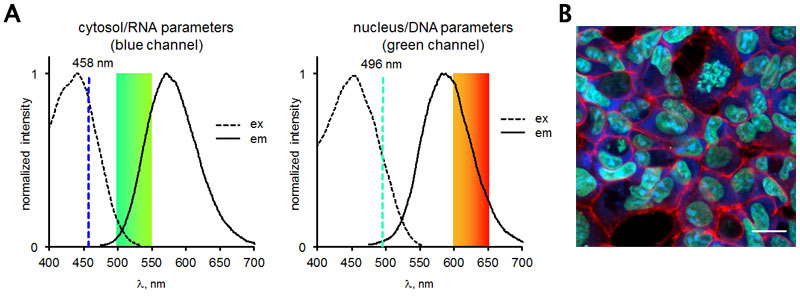
(A) Excitation and emission spectra of Miami Orange. (B) Representative image of cells (HEK293) treated with Miami Orange.
- Pitter DR, Brown AS, Baker JD, Wilson JN. One probe, two-channel imaging of nuclear and cytosolic compartments with orange and red emissive dyes. Org Biomol Chem. 2015 Aug 10. doi: 10.1039/C5OB01428J
- Ansari JM, Abraham NM, Massaro J, Murphy K, Smith-Carpenter J, Fikrig E. Anti-Biofilm Activity of a Self-Aggregating Peptide against Streptococcus mutans. Front Microbiol. 2017 Mar 24;8:488. doi: 10.3389/fmicb.2017.00488. eCollection 2017. View Article
- Quyen TL, Ngo TA, Bang DD, Madsen M, Wolff A. Classification of Multiple DNA Dyes Based on Inhibition Effects on Real-Time Loop-Mediated Isothermal Amplification (LAMP): Prospect for Point of Care Setting. Front Microbiol. 2019 Oct 15;10:2234. View article
If you publish research with this product, please let us know so we can cite your paper.