Anti-Osteopontin Antibodies

This range of mouse monoclonal antibodies was generated against purified recombinant human Osteopontin (OPN) and recognizes epitopes throughout OPN.
Osteopontin (OPN, also designated Bone Sialoprotein 1, Urinary Stone Protein, spp-1, eta-1, nephropontin, uropontin) is a phosphoglycoprotein synthesized in a variety of tissues and cells. It was originally identified as a bone matrix protein and subsequently as a cytokine produced by various immune and transformed cell lines. OPN is a biomarker for various types of cancers and inflammatory diseases. Excessive or dysregulated OPN expression has been linked to the pathogenesis of both autoimmune disorders such as multiple sclerosis, systemic lupus erythematosus, rheumatoid arthritis, atherosclerosis and other inflammatory diseases including cardiovascular disease, chronic obstructive pulmonary disease, inflammatory bowel disease, liver disease and asthma.
From the laboratory of David T. Denhardt, PhD, Rutgers University.
| Catalog Number | Product | DataSheet | Size | AVAILABILITY | Price | Qty |
|---|
| Product Type: | Antibody |
| Accession ID: | P10451 |
| Comments: | See "Comments" below for Specifications |
| Shipped: | Cold packs |
Not sure which antibody is right for your research? Try our Osteopontin Antibody Sampler Pack!
mAb recognition of OPN in Western Blots
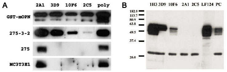
A: Western blots showing antibody recognition of murine OPN. Conditioned media (10 ul/lane) from various cell lines or 50 ng of recombinant murine OPN (GST-mOPN) were separated on 12% SDSPAGE gels and transferred to PVDF membranes. The membranes were then cut into strips which were blotted with monoclonal antibodies at 1 ug/ml or polyclonal control (shown above each lane). 275-3-2: ras-transformed murine fibroblast cell line. 275: non-transformed murine fibroblast 3T3 cell line. MC3T3E1: preosteoblast cell line induced to differentiate for 12 days. B: Western blot of human urine detected with anti-OPN mAbs. Urine was collected and dialyzed extensively against 0.1M NaCl before approximately tenfold concentration with Centriprep spin columns. Five microliters of the concentrated, dialyzed urine was assayed via SDSPAGE and Western blotting with mAbs at 1 ug/ml. Polyclonal antibody LF124 (kindly provided by Dr. Larry Fisher, NIH) and polyclonal anti-OPN (recombinant) mouse serum were used as controls.
Adapted from: Wang KX, et al. J Immunol. 2009 Feb 15;182(4):2485-91.
Antibody 2C5 blocks chronic restraint stress (CRS)-induced thymus atrophy in mice
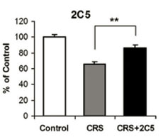
OPN+/+ mice in a BALB/c or 129 background were injected with the anti-OPN mAb 2C5 (100 ug) 24 h before CRS and immediately before each cycle of restraint. Animals were divided into three groups: control group (n = 24), untreated; CRS group (n = 56), injected with PBS and restrained; CRS + mAb group (n = 57) was injected with OPN in PBS and restrained. At the end of the CRS session (two 24-h cycles for BALB/c mice, three 24-h cycles for 129 mice), the thymi were evaluated for loss of weight compared with the control group. Data represent mean ± SEM. ??, p < 0.01.
Adapted from: Wang KX, et al. J Immunol. 2009 Feb 15;182(4):2485-91.
Antibody 3D9 inhibits cell adhesion to recombinant OPN
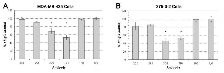
Tissue culture treated 96-well plates were coated with 150 uM human recombinant his-tagged OPN, then blocked with 1% BSA. Antibodies were then added at 125 uM and allowed to bind OPN for 2 h. The wells were then washed and 5 x 10⁴ MDA-MB-435 (A) or 275-3-2 (B) cells were added and allowed to adhere for 3 or 3.5 h respectively. Non-adherent cells were removed by washing and adherent cells were quantitated by staining with crystal violet. Data are representative of four independent experiments for the MDA-MB-435 cell line and two independent experiments for the 275-3-2 cell line (n=4). *P<0.001 Students t-test.
Adapted from: Kazanecki CC, et al. J Cell Biochem. 2007 Nov 1;102(4):925-35.
Anti-Osteopontin (OPN) Antibodies
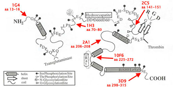
| Clone Name: | 1G4 | 1H3 | 2C5 | 2A1 | 10F6 | 3D9 | |
| Antigen Name: | Osteopontin (OPN) | Osteopontin (OPN) | Osteopontin (OPN) | Osteopontin (OPN) | Osteopontin (OPN) | Osteopontin (OPN) | |
| Acession ID: | P10451 | P10451 | P10451 | P10451 | P10451 | P10451 | |
| IgG Isotype: | IgG2a, kappa | IgG1, kappa | IgG1, kappa | IgG1, kappa | IgG1, kappa | IgG1, kappa | |
| Clonality: | Monoclonal | Monoclonal | Monoclonal | Monoclonal | Monoclonal | Monoclonal | |
| Species Reactivity: | Human | Human and mouse | Human and mouse | Human and mouse | Human and mouse | Human and mouse | |
| Epitope: | aa 13-16 | aa 70-80 | aa 141-151 | aa 206-208 | aa 225-272 | aa 298-315 | |
| Type of Immunogen: | Recombinant histidine-tagged human OPN (prepared in E. coli and purified on a nickel column) | ||||||
| Species Immunized: | Mouse | Mouse | Mouse | Mouse | Mouse | Mouse | |
| Purification Method: | Protein G | Protein G | Protein G | Protein G | Protein G | Protein G | |
| Amount: | 50ug | 50ug | 50ug | 50ug | 50ug | 50ug | |
| Storage Buffer: | 0.1M Sodium Phosphate, pH 7.4, 0.15M NaCl, 0.05% (w/v) Sodium Azide | ||||||
| Tested Applications: | WB, ELISA, IP, IHC | WB, ELISA, IP, Blocking | WB, ELISA, IP, IHC, Blocking | WB, ELISA, IP, IF, IHC | WB, ELISA, IP | WB, ELISA, IP | |
| Storage Temperature: | -20C | -20C | -20C | -20C | -20C | -20C | |
| Shipped: | Cold packs | Cold packs | Cold packs | Cold packs | Cold packs | Cold packs | |
| Comments: | Binds strongly to all four human forms of OPN | Increased affinity towards O-glycosylated OPN. | Recognizes both native and post-translationally modified forms of OPN. | Inhibits cell adhesion to OPN by 40-50% in MDA-MB-435 breast cancer cells and 275-3-2 murine ras-transformed fibroblast cells | |||
- Kazanecki CC, Kowalski AJ, Ding T, Rittling SR, Denhardt DT. Characterization of anti-osteopontin monoclonal antibodies: Binding sensitivity to post-translational modifications. J Cell Biochem. 2007 Nov 1;102(4):925-35.
- Wang KX, Denhardt DT. Osteopontin: role in immune regulation and stress responses. Cytokine Growth Factor Rev. 2008 Oct-Dec;19(5-6):333-45.
- Christensen B, Klaning E, Nielsen MS, Andersen MH, Sorensen ES. C-terminal Modification of Osteopontin Inhibits Interaction with the ?V?3-Integrin. J Biol Chem. 2012 February 3; 287(6): 37883797.
- Wang KX, Shi YF, Ron Y, Kazanecki CC, Denhardt DT. Plasma osteopontin modulates chronic restraint stress-induced thymus atrophy by regulating stress hormones: inhibition by an anti-osteopontin monoclonal antibody. J Immunol. 2009 Feb 15;182(4):2485-91.
- Nagao M, Feinstein TN, Ezura Y, Hayata T, Notomi T, Saita Y, Hanyu R, Hemmi H, Izu Y, Takeda S, Wang K, Rittling S, Nakamoto T, Kaneko K, Kurosawa H, Karsenty G, Denhardt DT, Vilardaga JP, Noda M. Sympathetic control of bone mass regulated by osteopontin. Proc Natl Acad Sci U S A. 2011 Oct 25;108(43):17767-72.
If you publish research with this product, please let us know so we can cite your paper.