Anti-Lamin A+C [WL4G10] Antibody
![Anti-Lamin A+C [WL4G10] Antibody](https://www.kerafast.com/MediaStorage/Product/Images/Large/1758_2001202001270017570.jpg "Anti-Lamin A+C [WL4G10] Antibody")
This mouse IgG1 kappa monoclonal antibody was raised against Glutathione-S-transferase fused to C-terminal fragment of human lamin A (amino acids residues 422-664) and recognizes human lamins A and C.
Highlights:
- Reacts with human Lamins A and C
- Suitable for Western Blot, Immunohistochemistry, Immunocytochemsitry and Immunofluorescence applications
Lamins A and Care major components of the nuclear lamina, a thin protein meshwork located at the nuclear face of the nuclear envelope (NE). The lamina provides structural integrity to the NE and is involved in many other aspects of nuclear biology including transcription and chromatin organization. Lamins A and C arise from the LMNA gene by alternative splicing and the majority of adult tissues express at least one of the isoform. The LMNA gene has been extensively studies due to its association with variety of human diseases. Mutations in LMNA have been linked 12 distinct disorders, including Emery-Dreifus muscular dystrophy, dilated cardiomyopathy, Dunnigan-type partial lipodystrophy and Hutchinson-Gilford progeria syndrome.
From the laboratory of Brian Burke, PhD, A*STAR Institute of Medical Biology (IMB).
| Product Type: | Antibody |
| Accession ID: | PO2545 |
| Antigen: | Lamins A and C |
| Molecular Weight: | Lamin A 74 kDa, Lamin C 65 kDa |
| Isotype: | IgG1k |
| Clonality: | Monoclonal |
| Clone Name: | WL4G10 |
| Reactivity: | Human, does not recognize mouse lamins A+C, not tested with other species |
| Immunogen: | Glutathione-S-transferase fused to C-terminal fragment of human lamin A (amino acids residues 422-664) |
| Species Immunized: | Mouse |
| Buffer: | Cell Culture Supernatant |
| Positive Control: | Hela, 293T |
| Tested Applications: | WB, IHC, ICC, IF |
| Comments: | Knock-in validated |
| Storage: | -80C |
| Shipped: | Cold Packs |
Western Blot
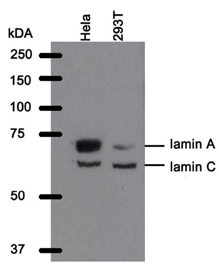
Western blot analysis of WL4G10 antibody. Whole cell lysate from He/a cells and 293T cells were separated on SOS-PAGE gel, transferred to nitrocellulose membrane and stained with WL4G10 antibody. The membrane was blocked overnight in blocking buffer {5% non-fat dry milk resuspended in 0.1% Tween and 1X PBS). Both primary and secondary antibodies were diluted in blocking buffer. As expected 293T cells express less of the Jamin A isoform.
Confocal images of human Hela cell lines immunofluorescently stained for lamin A+C with WL4G10 antibody.
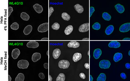
The WL4G10 antibody works on cells fixed with 4% PFA (upper panel) and cold methanol (lower panel). Nuclei were counterstained with Hoechst. The antibody is not suitable to detect mouse lamin A+C.
The mouse monoclonal WL4G10 antibody recognizes human lamins A and C.
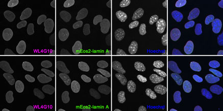
Immortalized mouse adult LMNA null fibroblast stably expressing mEos2-lamin A (upper pnale) or mEos2- lamin C (2) (bottom panel) were fixed in 4% PFA and stained with WL4G10 antibody (magenta). Expression of the respective LMNA isoform is shown in green. The nuclei were counter-stained with Hoechst. Of note the WL4G10 antibody does not recognize mouse lamins A and C.
Immunofluorescence analysis of paraffin-embedded human gut with WL4G10 antibody.
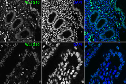
Following heat-induced antigen retrieval (Dako antigen retrieval solution pH6 or pH9) the sections were stained with mouse monoclonal anti-lamin A+C clone WL4G10 antibody (green). Nuclei were visualized with DAPI.
- Burke, B., C.L Stewart. 2013. The nuclear lamins: flexibility in function. Nat Rev Mol Cell Biol. 14(1):13-24.
- Xie, W., A. Chojnowski, T. Boudier, J. S. Lim, S. Ahmed, Z. Ser, C. Stewart, B. Burke. 2016. A-type Lamins Form Distinct Filamentous Networks with Differential Nuclear Pore Complex Associations. Curr Biol. 2016 Sep 9. pii: S0960-9822(16)30842-9. doi: 10.1016/j.cub.2016.07.049.
If you publish research with this product, please let us know so we can cite your paper.