Goat Anti-Trem2 (Mouse) Antibody
 Antibody")
This goat IgG polyclonal antibody was generated against peptide sequence CQVEHSTSRNQET from the internal region of triggering receptor expressed on myeloid cells 2 (Trem2) and recognizes mouse Trem2.
Highlights:
- Reacts with mouse Trem2
- Suitable for Peptide ELISA and Immunofluorescence applications
- Mutations in the Trem2 gene are thought to increase the risk of Alzheimer's disease
Triggering receptor expressed in myeloid cells 2 (Trem2) is a pattern recognition receptor found on the plasma membrane of myeloid cells which include macrophages, granulocytes, monocytes, and dendritic cells. When activated, Trem2 induces an innate immune response that includes phagocytosis, chemotaxis, and transcriptional changes. Consequently, Trem2 may be involved in chronic inflammation. Within the brain, Trem2 is expressed by microglia and is upregulated on microglia around amyloid plaques in Alzheimer's disease. Mutations in the Trem2 gene are thought to increase the risk of Alzheimer's disease.
| Product Type: | Antibody |
| Name: | Goat Anti-Trem2 (Mouse) Antibody |
| Alternative Name(s): | Triggering receptor expressed on monocytes 2, Trem2a, Trem2b, Trem2c |
| Accession ID: | NP_112544.1 |
| Antigen: | Trem2 (Mouse) |
| Isotype: | IgG |
| Clonality: | Polyclonal |
| Reactivity: | Mouse |
| Specificity: | Trem2 (Mouse) |
| Immunogen: | CQVEHSTSRNQET |
| Species Immunized: | Goat |
| Epitope: | Internal Region |
| Purification Method: | Purified from goat serum by ammonium sulphate precipitation followed by antigen affinity chromatography using the immunizing peptide |
| Buffer: | Supplied at 0.5 mg/ml in Tris saline, 0.02% sodium azide, pH7.3 with 0.5% bovine serum albumin. |
| Tested Applications: | Pep-ELISA, IF |
| Storage: | Aliquot and store at -20C. Minimize freezing and thawing. |
| Shipped: | Cold Packs |
Immunofluorescence
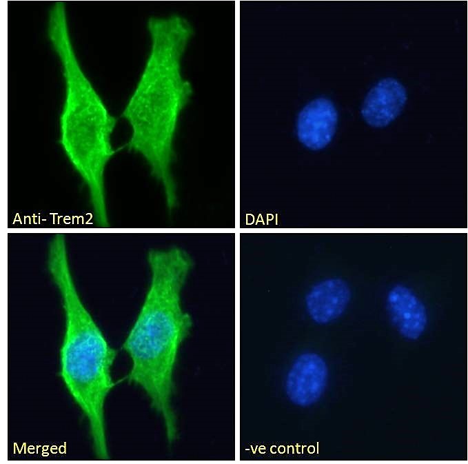
Immunofluorescence analysis of paraformaldehyde fixed 3T3-L1 cells, permeabilized with 0.15% Triton. Primary incubation 1hr (10ug/ml) followed by Alexa Fluor 488 secondary antibody (2ug/ml), showing membrane/cytoplasmic staining. The nuclear stain is DAPI (blue). Negative control: Unimmunized goat IgG (10ug/ml) followed by Alexa Fluor 488 secondary antibody (2ug/ml).
If you publish research with this product, please let us know so we can cite your paper.