Goat Anti-ZO-1 Antibody

This goat IgG polyclonal antibody was generated against peptide sequence C-PKPTSQNQFSEHDKT from the internal region of Zonula occludens-1 (ZO-1) and recognizes human ZO-1.
Highlights:
- Reacts with human ZO-1
- This antibody is expected to recognize both reported isoforms (NP_003248.3; NP_783297.2)
- Suitable for Peptide ELISA and Immunofluorescence applications
Zonula occludens-1 (ZO-1), or Tight junction protein-1 (TJP1), is a peripheral membrane protein that belongs to the family of zonula occludens proteins. Of these tight junction-associated proteins, Zo-1 was the first to be cloned. Its role includes serving as a scaffold protein that links the tight-junction transmembrane proteins to the actin cytoskeleton, and may also be involved in signal transduction at cell-cell junctions. This gene may also be associated with several diseases, such as Brain Edema and Corneal Endothelial Dystrophy.
| Product Type: | Antibody |
| Name: | Goat Anti-ZO-1 Antibody |
| Alternative Name(s): | TJP1 |
| Accession ID: | NP_003248.3; NP_783297.2 |
| Antigen: | ZO-1 |
| Isotype: | IgG |
| Clonality: | Polyclonal |
| Reactivity: | Human |
| Specificity: | ZO-1 (Expected to recognize both reported isoforms NP_003248.3 & NP_783297.2.) |
| Immunogen: | CPKPTSQNQFSEHDKT |
| Species Immunized: | Goat |
| Epitope: | Internal region |
| Purification Method: | Purified from goat serum by ammonium sulphate precipitation followed by antigen affinity chromatography using the immunizing peptide |
| Buffer: | Supplied at 0.5 mg/ml in Tris saline, 0.02% sodium azide, pH7.3 with 0.5% bovine serum albumin. |
| Tested Applications: | Pep-ELISA, IF |
| Storage: | Aliquot and store at -20C. Minimize freezing and thawing. |
| Shipped: | Cold Packs |
Immunofluorescence
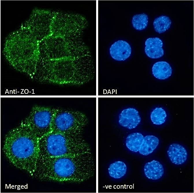
Immunofluorescence analysis of paraformaldehyde fixed A431 cells, permeabilized with 0.15% Triton. Primary incubation 1hr (10ug/ml) followed by Alexa Fluor 488 secondary antibody (2ug/ml), showing cellular junction staining. The nuclear stain is DAPI (blue). Negative control: Unimmunized goat IgG (10ug/ml) followed by Alexa Fluor 488 secondary antibody (2ug/ml).
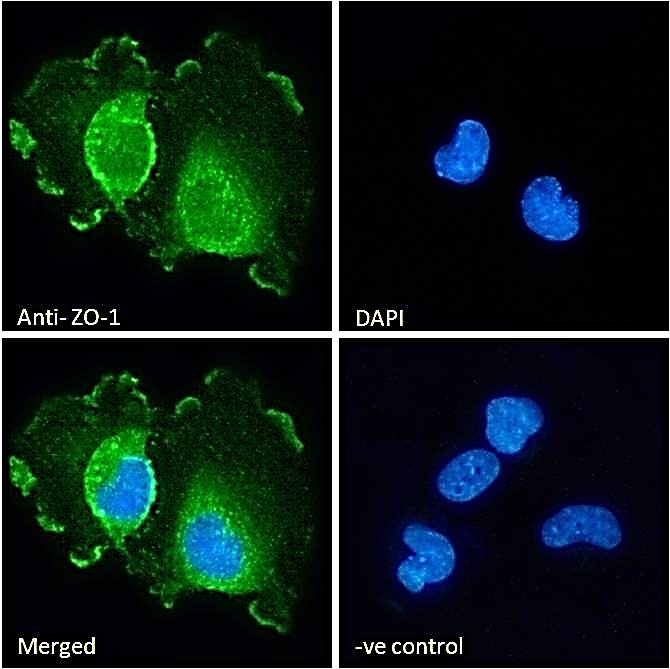
Immunofluorescence analysis of paraformaldehyde fixed U251 cells, permeabilized with 0.15% Triton. Primary incubation 1hr (10ug/ml) followed by Alexa Fluor 488 secondary antibody (2ug/ml), showing plasma membrane and cytoplasmic staining. The nuclear stain is DAPI (blue). Negative control: Unimmunized goat IgG (10ug/ml) followed by Alexa Fluor 488 secondary antibody (2ug/ml).
If you publish research with this product, please let us know so we can cite your paper.