GlowDot10 Protein-Based Fluorescent Nanoparticles

Single protein nanoparticles that retain the structure and function of native protein used.
Highlights:
- Tunable size and color over nm level or better
- Particle surface is rich in COOH groups for additional conjugation or manipulations
- Exhibits superior thermal stability to native proteins, as tested with steam sterilization studie
- Metal ion-free - likely to be non-toxic to most cells
From the laboratory of C. Vijaya Kumar, PhD, University of Connecticut.
| Catalog Number | Product | DataSheet | Size | AVAILABILITY | Price | Qty |
|---|
Specifications
| Product Type: | Small Molecule |
| Internal Contents: | Protein, lipid, carbohydrate and buffer salts |
| Functionalization: | Abundant surface COOH groups |
| Particle Morphology: | Nearly spherical |
| Format: | Powder containing sucrose or glucose |
| Concentration: | 25% solid in 75% sucrose or glucose |
| Spectral Information: |
GlowDot10-350 - Ex: 350nm, Em: 410nm GlowDot10-425 - Ex: 425nm, Em: 460nm GlowDot10-494 - Ex: 494nm, Em: 525 GlowDot10-580 - Ex: 580nm, Em: 605nm GlowDot10-365W - Ex: 365nm |
| Size Distribution: | ±2nm |
| Mean Diameter: | 10 nm |
| Nanoparticle Consistency: | >90% |
| Comments: |
Refractive index = 1.33 Zeta potential = -23mV |
| Storage: | -20C |
| Shipped: | Cold packs |
Data
GlowDot10 Characterization

(Left) Overlay of the circular Dichroism spectra of GlowDot10-N (red line) and unmodified bovine serum albumin (black), confirming that the secondary structure of the protein is completely retained after particle synthesis. (Center) Dynamic light scattering showing the average hydrodynamic size of 13 nm. (Right) TEM (scale bar 100 nm) showing 10 nm sized particles.
GlowDot10 Absorbance Spectra
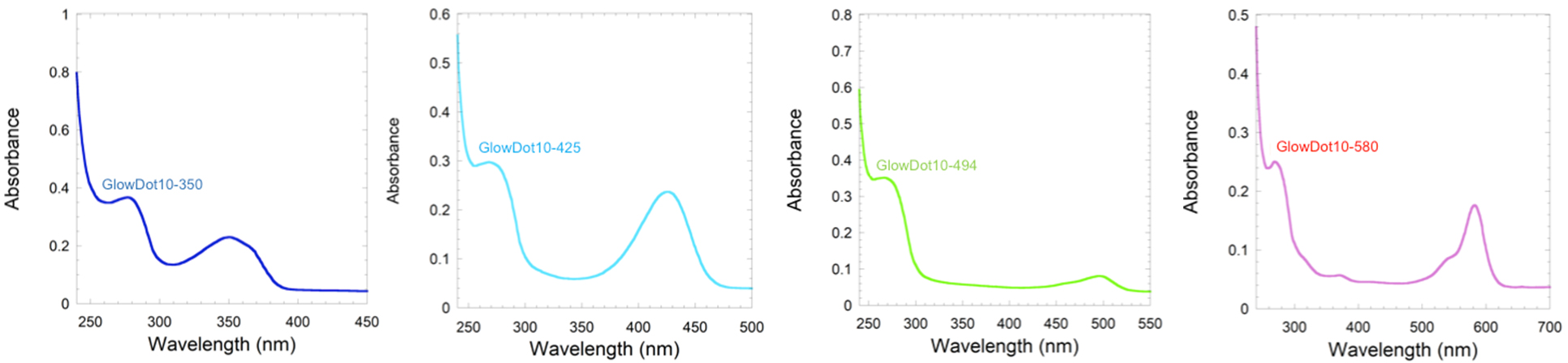
(Left to right) GlowDot10-350, GlowDot10-425, GlowDot10-494, GlowDot10-580
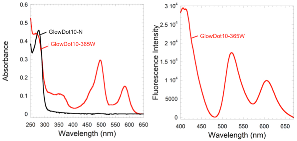
(Left) Absorbance spectra of GlowDot10-N (black line) compared to GlowDot10-365W (red line). (Right) Fluorescence spectrum of GlowDot10-365W (red line).
Provider
From the laboratory of C. Vijaya Kumar, PhD, University of Connecticut.
Comments
Recommended Protocol
- Remove centrifuge tube from packaging.
- Re-suspend centrifuge tube contents in 10 mM Phosphate buffer pH 7.2 so that the final volume is 300 uL. This will yield a 10 mg/mL solution of GlowDots.
- Filter re-suspended GlowDots using a 13 mm diameter PVDF filter with 0.22 µm pore size.
- Incubate cells with 0.3 mg/mL for 1 hour.
- Image cells on confocal microscope with appropriate excitation laser.
References
If you publish research with this product, please let us know so we can cite your paper.