Neuro-2a Split Superfolder GFP-Tau Cell Line (N2a-ssGT)
")
This mouse N2a-ssGT (Neuro-2a Split Superfolder GFP-Tau) Cell Line was derived from Neuro-2a cells. It was designed for measuring human tau dimers/oligomers in the split GFP-Tau oligomerization assay, thus serving as a useful tool for studying pathologies and dementias of the nervous system (e.g., Pick Disease, Alzheimer's Disease).
Highlights:
- Over-expresses GFP-Tau (two split super-folder GFP fragments)
- Co-transfected with both pmGFP10C-Tau and pmGFP11C-Tau plasmids
Microtubule associated protein tau (MAPT) forms dimers/oligomers and progressively aggregate into neurofibrillary tangles to cause neuronal cell death in diseases of tauopathy. Oligomeric tau species were crucial to induce tau transmission between neurons and toxicity within neurons. Mouse Neuro-2a cell line was engineered to stably over-express two split super-folder GFP fragments (1-212 a.a. of beta strands 1-10 and 213-228 a.a. of the 11th beta-strand) that both fused with a monomeric human wild-type tau protein (4R0N). Dimerization of tau molecules simultaneously induces GFP-fragment complementation and fluorescence. This novel established technique allows for both visualization and quantification of tau dimerization/oligomerization based on the green fluorescent protein (GFP)-fragment complementation. Furthermore, the process of tau dimerization/oligomerization can be measured by detecting green fluorescence, providing a valuable tool for investigating tau oligomer modifying drugs and treatments.
From the laboratory of Daniel C Lee, PhD and Chao Ma, University of South Florida.
| Product Type: | Cell Line |
| Name: | Neuro-2a Split Superfolder GFP-Tau (N2a-ssGT) |
| Cell Type: | Neuroblast, adherent |
| Accession ID: | P10636 |
| Morphology: | Neuronal and amoeboid stem cells |
| Source: | Brain |
| Species: | Mus musculus, mouse |
| Biosafety Level: | BSL-1 |
| Subculturing: | Subculture at a ratio of 1:5 or 1:10 using 0.05% Trypsin-EDTA at 2.0 ml per 75 cm2 flask at 37 °C incubator for 5 minutes. |
| Growth Conditions: | DMEM (Gibco, #11965167, 1X), 10% (v/v) fetal bovine serum (Sigma-Aldrich, #12306C), 1% (v/v) GlutaMAX (Gibco, #35050061, 100X), 1% (v/v) MEM Non-Essential Amino Acids Solution (NEAA, Gibco, #11140050, 100X), 1% (v/v) Penicillin-Streptomycin (10,000 U/mL, Gibco, #15140122, 100X), 1% (v/v) Sodium Pyruvate (Gibco, # 11360070, 100 mM, 100X), 125 ug/ml Zeocin (Gibco, #R25001, 100 mg/ml), 400 ug/ml G418 sulfate (Corning, #30234CI, 50 mg/ml). Please also see links below under "Documentation." |
| Cryopreservation: | Recovery™ Cell Culture Freezing Medium (Gibco, #12648010) or Complete Growth Medium supplemented with 10% (v/v) DMSO |
| Storage: | LN2 |
| Shipped: | Dry Ice |
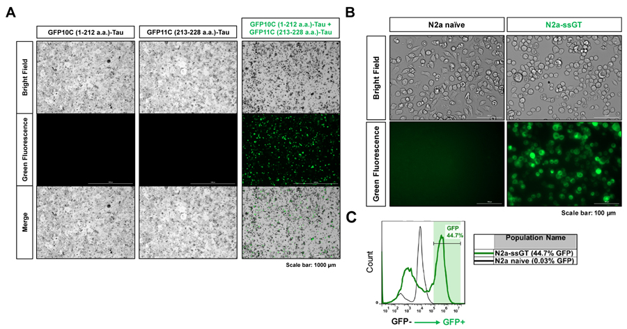
(A) Representative images of Neuro-2a cells transfected with different combination of split GFP-Tau plasmids. (B) Representative images of a stable single clone of N2a-ssGT cell line. (C) Measurement of GFP positive percentage in N2a-ssGT by flow cytometry.
Adapted from: Sandusky-Beltran LA, et al. J Clin Invest. 2021;131(4).
To develop a stable monoclonal cell line, mouse Neuro-2a cells (N2a, ATCC®, #CCL131™) were co-transfected with both pmGFP10C-Tau (Addgene, #71433) and pmGFP11C-Tau (Addgene, #71434) plasmids. Then transfected cells were replaced with N2a growth medium containing zeocin (125 µg/ml) and G418 (400 µg/ml) for selection of stable cell clones. After a long-term selection process (up to 6 months), several single clones were picked by cloning cylinder (Sigma-Aldrich, #Z370789) and eventually proliferated into stable monoclonal cell lines. To characterize the new cell lines, the cells were imaged for GFP by using Cytation™ 3 Cell Imaging Multi-Mode Reader (BioTek™). GFP expression was also measured using flow cytometry to confirm that cells maintained a relatively stable high GFP positive percentage using Accuri® C6 flow cytometer (BD Biosciences) under FL1 detector (Laser 488).
- Sandusky-Beltran LA, Kovalenko A, Ma C, Calahatian JIT, Placides DS, Watler MD, et al. Spermidine/spermine-N(1)-acetyltransferase ablation impacts tauopathy-induced polyamine stress response. Alzheimers Res Ther. 2019;11(1):58.
- Sandusky-Beltran LA, Kovalenko A, Placides DS, Ratnasamy K, Ma C, Hunt JB, Jr., et al. Aberrant AZIN2 and polyamine metabolism precipitates tau neuropathology. J Clin Invest. 2021;131(4).
If you publish research with this product, please let us know so we can cite your paper.