Anti-AP-Tag [EF10] Antibody
![Anti-AP-Tag [EF10] Antibody](https://www.kerafast.com/MediaStorage/Product/Images/Large/1046_2001202001263210450.jpg "Anti-AP-Tag [EF10] Antibody")
This mouse IgG1k monoclonal antibody was raised against a synthetic peptide (GLNDIFEAQKIEWHE) and recognizes biotinylable peptide (Epitope-Tag) called AP-tag .
Highlights:
- Biotin-based detection system, applicable to the analysis of transmembrane protein trafficking (i.e., quantification of receptor-internalization from the cell surface into the cell)
- Binding of EF10 to the target protein is not affected by biotinylation of the acceptor sequence
- Suitable for Immunofluorescence staining, ELISA, Biological Function (Endocytosis Assay) and Flow Cytometry applications
Epitope tags have been widely used in the study of protein expression in various systems. The 15-residue peptide served as a substrate mimic for biotin ligase (BirA), which usually recognizes the much larger protein domain. Using this antibody an alternative method was developed to analyse receptor internalization and recycling which allows a more detailed analysis of receptor trafficking compared to classical antibody-based detection methods. This biotin-based detection system may be generally applicable to the analysis of transmembrane protein trafficking.
From a laboratory at University of Göttingen.
| Product Type: | Antibody |
| Antigen: | AP-tag, GLNDIFEAQKIEWHE |
| Isotype: | IgG1k |
| Fusion Tag(s): | X63Ag8.653 myeloma cells |
| Clonality: | Monoclonal |
| Clone Name: | EF10 |
| Specificity: | AP-tag, GLNDIFEAQKIEWHE |
| Immunogen: | GLNDIFEAQKIEWHEC-KLH |
| Species Immunized: | Mouse |
| Purification Method: | Protein G |
| Buffer: | PBS, 0.05% (w/v) Sodium Azide |
| Tested Applications: | Immunofluorescence staining, ELISA, Biological Function (Endocytosis Assay), Flow Cytometry |
| Storage: | -20C |
| Shipped: | Cold packs |
Double immunoflourescence (anti-AP/streptavidin) of CXCR4-/CCR5-expressing cells during ligand- induced internalization (30’) and recycling (60’)
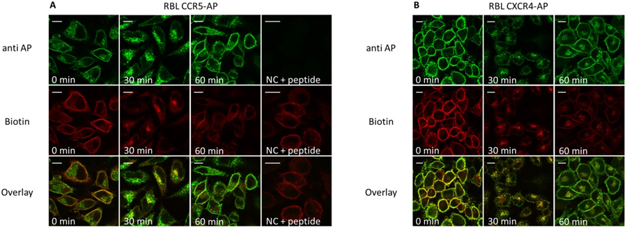
Prior to stimulation and staining CXCR4-AP (right) and CCR5-AP (left) expressing RBL cells were seeded on glass cover slips. Cells were enzymatically biotinylated and stimulated with the corresponding ligand (125 nM CCL5, CXCL12; 30’/37°C). Recycling was induced by acid wash and transfer into ligand-free medium containing receptor antagonists (30 μM AMD 3100; 3 μM TAK779). Cells were fixed with 3% PFA (15’/37°C) and permeabilized with 0.1% saponin (15’/37°C). 10 μg/ml anti-AP antibody (anti AP; with or without preincubation with 2mg /ml of the AP-peptide) and 2 μg/ml streptavidin-Alexa647 (Biotin) was used for staining (60’/on ice/dark). Samples were fixed with mounting medium and analyzed by confocal laser scanning microscopy. Scale bar 10 μM.
Adapted from: Liebick M, Schläger C, Oppermann M (2016) PLOSOne 11(6):e0157502.
- Liebick M, Schläger C, Oppermann M (2016) Analysis of Chemokine Receptor Trafficking by Site-Specific Biotinylation. PLOSOne 11(6):e0157502.
If you publish research with this product, please let us know so we can cite your paper.