Human Endometrial Stromal Cell Line (SHT290)
")
Cell line SHT290 derived by introduction of the human telomerase reverse transcriptase (hTERT) into normal human endometrial stromal cells.
Highlights:
- Cells function like normal endometrial stromal according to criteria of proliferation, karyotype, cellular localization of cytoskeletal markers and nuclear staining, and basal gene expression based on microarray analysis
Endometrial stromal cells are the main supportive (stromal) cell type that underlies endometrial surface epithelium and surrounds glands. In the human endometrium, stromal cells mediate the proliferative response of epithelial cells to the steroid hormones estrogen and progesterone. The development and characterization of these cells was reported in a publication: (See: Barbier, CS et al., Biol Reprod. 2005 Jul;73(1):106-14.). These cells function like normal human endometrial stromal cells in their support of normal endometrial epithelial cell function in vitro that mimics the functions of the epithelial cells in vivo.
From the laboratory of David G. Kaufman, MD, PhD, University of North Carolina Chapel Hill.
| Product Type: | Cell Line |
| Cell Type: | Endometrial Stromal Cell |
| Accession ID: | CVCL_G213 |
| Morphology: | Depends on growth conditions, bipolar to polygonal |
| Source: | Endometrium |
| Organism: | Human |
| Biosafety Level: | BSL 2 |
| Subculturing: | Subculture at no higher a split ratio than one-to-three in order to preserve high culture density. Monitor trypsinization visually to prevent over-trypsinization. Quench tripsin with medium containing 10% serum. |
| Growth Conditions: | Use SM medium consisting of a 1:1 mixture of Ham F12 (Gibco, Invitrogen Corp., Carlsbad, CA) and M199 basic medium (Sigma, St. Louis, MO) supplemented with 5% bovine calf serum (BCS, Hyclone, Logan, UT), 0.1% Mitoplus (BD Biosciences, Becton Dickinson, Franklin Lakes, NJ), and 2 ug/ml of insulin (Sigma) and antibiotic/antimycotic solution diluted to yield 100 units/ml penicillin G sodium, 100 mg/ml streptomycin sulfate and 250 ng/ml amphotericin B (ABAM, Gibco). This medium allows for co-culture with human endometrial epithelial cells. |
| Cryopreservation: | 20 % FBS, 10% DMSO, M199/F12 Medium |
| Cell Passage Number: | 64 |
| Comments: | Cells grow slowly and must be maintained at high density. Plate cells initially 30 mm dish or in well of a six-well dish. Expand into larger dishes as the number of cells increases. These cells are very fastidious. Follow culturing instructions carefully to successfully propagate cells. |
| Storage: | LN2 |
| Shipped: | Dry Ice |
Comparative cell characterization of endometrial cells
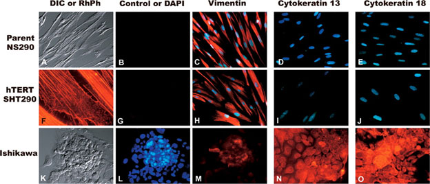
The presumptive mesenchymal cell identity of SHT290 telomerized stromal cells was compared to parent NS290 cells. Ishikawa cells were used as a positive control for epithelial cell markers. Column with A, F, and K: (A) Differential interference contrast (DIC) image of parent NS290 stromal cells showing their typical fibroblast-like bipolar morphology; (F) SHT290 cells labeled with rhodamine-phalloidin show details of their filamentous actin cytoskeleton, stress fibers, and intercellular contacts; (K) DIC image of Ishikawa cells showing their epithelial growth pattern. B and G are controls for rhodamine-labeled secondary antibodies used for all other cytoskeletal markers. L shows typical nuclear staining of Ishikawa cells with Hoechst stain. Blue in any image indicates Hoechst-labeled nuclear DNA. The pattern of vimentin labeling (red) in all three cell types is shown in the column with C, H, and M; note that the telomerized derivative SHT290 cells (H) exhibit the strong vimentin expression of their NS290 parent cells (C). Cytokeratin profiles of stromal cells double-labeled for DNA are shown in D and I for cytokeratin 13 and in E and J for cytokeratin 18; note the negligible red signal for cytokeratins in the stromal phenotype. Conversely, the fluoroprobe signals for cytoskeletal expression markers of cytokeratin 13 (N) and cytokeratin 18 (O) were strong and predominantly cytoplasmic in Ishikawa cells of epithelial origin. Magnification ×200 (A–E and G–O) and ×630 (F).
Adapted from: Barbier, CS et al., Biol Reprod. 2005 Jul;73(1):106-14.- Barbier CS, Becker KA, Troester MA, Kaufman DG. Expression of exogenous human telomerase in cultures of endometrial stromal cells does not alter their hormone responsiveness. Biol Reprod. 2005 Jul;73(1):106-14. Epub 2005 Mar 16. PubMed PMID: 15772261.
- Barbier C, Kloosterboer HJ, Kaufman DG. Effects of tibolone metabolites on human endometrial cell lines in co-culture. Reprod Sci. 2008 Jan;15(1):75-82.
- Smalley T, Metcalf R, Patel R, Islam SMA, Bommareddy RR, Acevedo-Duncan M. The Atypical Protein Kinase C Small Molecule Inhibitor ζ-Stat, and Its Effects on Invasion Through Decreases in PKC-ζ Protein Expression. Front Oncol. 2020 Feb 27;10:209. View article
If you publish research with this product, please let us know so we can cite your paper.