Anti-Exocyst Complex, Sec5 (EXOC2) Subunit [5D8A10] Antibody
 Subunit [5D8A10] Antibody")
This monoclonal antibody recognizes exocyst complex sec5 subunit (EXOC2) and is reactive with mouse and rat.
Exocyst complex (also known as the sec6/8 complex) is a multimeric complex that has been implicated to play a role in exocytosis. This complex is conserved from yeast to mammals and is composed of eight subunits (Sec3, Sec5, Sec6, Sec8, Sec10, Sec15, Exo70, and Exo84). In the budding yeast Saccharomyces cerevisiae, mutations in these complex subunits promote cytoplasmic accumulation of secretory vesicles and affects polarized growth. In cultured Drosophila and mammlian cells, disruption of exocyst subunit function has been found to decrease neurite outgrowth and Golgi-to-plasma membrane vesicle trafficking. Furthermore, a delay in neuronal induction was observed in mouse embryos with deletion of the sec8 gene, suggesting that the exocyst complex may play a role in cell differentiation and polarized growth.
From the laboratory of Shu-Chan Hsu, PhD, Rutgers University.
| Product Type: | Antibody |
| Name: | Anti-exocyst sec5 subunit (5S8A10) monoclonal antibody |
| Accession ID: | Q96KP1 |
| Host: | Mouse |
| Isotype: | IgG subtype not characterized |
| Clonality: | Monoclonal |
| Clone Name: | 5D8A10 |
| Specificity: | This antibody recognizes exocyst complex sec5 subunit. Demonstrated to react with rat and mouse tissues. |
| Immunogen: | Recombinant protein corresponding to full-length rat brain sec5 subunit |
| Format: | Liquid |
| Purification Method: | Protein G purified |
| Tested Applications: | Western blotting, ELISA and Immunofluorescence microscopy. |
| Concentration: | 1mg/mL |
| Amount: | 100uL |
| Storage: | Store at -20C |
| Shipped: | Cold packs |
Alternative names - Exoc2, mSec5
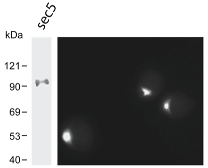
(left) Western blot analysis: 10 ug Rat brain lysate was resolved on a 8% SDS-polyacrylamide gel, transferred to nitrocellulose membrane and probed with 0.01ug/ml antibody.
(right) Immunofluorescence microcopy: Methanol-fixed neuroendocrine PC12 cells were stained with 0.1ug/ml antibody. For best visualization, the cells were incubated with the primary antibody at 4 OC overnight.
- Wang L, Li G, Sugita S. (2004) RalA-exocyst interaction mediates GTP-dependent exocytosis. J Biol Chem. 279:19875-19881.
- Fukai S, Matern HT, Jagath JR, Scheller RH, Brunger AT (2003) Structural basis of the interaction between RalA and Sec5, a subunit of the sec6/8 complex. EMBO J. 22:3267-78.
- Sugihara K, Asano S, Tanaka K, Iwamatsu A, Okawa K, Ohta Y (2002) The exocyst complex binds the small GTPase RalA to mediate filopodia formation. Nat Cell Biol. 4:73-8.
- Miao Y, Wu J, Abraham SN. Ubiquitination of Innate Immune Regulator TRAF3 Orchestrates Expulsion of Intracellular Bacteria by Exocyst Complex. Immunity. 2016 Jul 19;45(1):94-105. View Article
- Bhalla M, Van Ngo H, Gyanwali GC, Ireton K. The Host Scaffolding ProteinFilamin A and the Exocyst Complex Control Exocytosis during InlB-Mediated Entry of Listeria monocytogenes. Infect Immun. 2018 Dec 19;87(1). pii: e00689-18. View Article
If you publish research with this product, please let us know so we can cite your paper.