FITC-Trehalose

FITC-trehalose selectively labels live mycobacteria in vitro and in vivo, allowing for downstream fluorescent imaging of Mycobacterium tuberculosis in culture and in infected macrophages.
Highlights:
- Trehalose moiety - selectively incorporates into M. tuberculosis via the extracellular Ag85 enzymes
- FITC moiety - allows for simple downstream fluorescent imaging
- Shows no significant inhibition of M. tuberculosis growth - allows for imaging of healthy, viable bacteria
Tuberculosis is an infection that has plagued mankind for millennia, and remains a leading cause of death worldwide. A substantial obstacle to the development of new diagnostics, drugs and vaccines is the lack of tuberculosis-specific probes that can be used to rapidly assess infection and monitor response to treatment.
From the laboratory of Benjamin G. Davis, PhD, University of Oxford.
| Product Type: | Small Molecule |
| Name: | FITC-Trehalose |
| Alternative Name(s): | 1-Deoxy-α-D-gluco-hept-2-ulopyranosyl-(2→1)-2-deoxy-2-(N'-(fluorescein-5-yl)-thioureido)-α-D-glucopyranoside |
| Chemical Formula: | C34H36N2O15S |
| Molecular Weight: | 744.72 |
| Format: | yellow solid |
| Purity: | 95+% (NMR, HPLC) |
| Solubility: | Soluble in MeOH and water |
| Spectral Information: |
[α]D = 72.2 (c = 0.18, MeOH); 1H NMR (500 MHz, D2O): δ 1.48 (3 H, s, C-1’), 3.26 (1 H, d, J3,4 = 9.8 Hz, H-3’), 3.49 (3 H, m, H4’, H-5’, H-4), 3.74 (6 H, m, H5, H-6’, H-6a, H-6b, H-7a’, H-7b’), 4.00 (1 H, at, J2,3 = J3,4 = 9.8 Hz, H-3), 4.41 (1 H, dd, J2,3 = 10.9 Hz, |
| Storage: | -20C, protect from light |
| Shipped: | Room temperature |
Labeling of individual bacilli in vitro and within infected macrophages
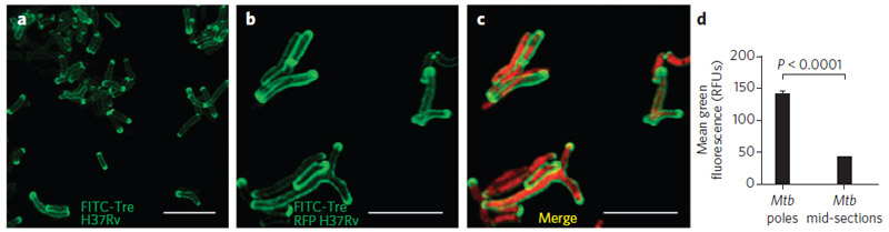
(a) H37Rv M. tuberculosis are labeled with FITC-trehalose and show signification accumulation of probe at the bacterial poles and membrane. (b) FITC-trehalose labeling of the RFP-expressing strain of H37Rv. (c) A merge of the FITC-trehalose and the RFP channels. (d) The mean fluorescence of poles and midsections of H37Rv M. tuberculosis, labeled with FITC-trehalose, are quantified and show statistically significant differential labeling. RFU, relative fluorescence units.
Adapted from: Backus, KM et al. Nat. Chem. Biol. 7, 228–235 (2011).
- Backus KM, Boshoff HI, Barry CS, Boutureira O, Patel MK, D'Hooge F, Lee SS, Via LE, Tahlan K, Barry CE 3rd, Davis BG. Uptake of unnatural trehalose analogs as a reporter for Mycobacterium tuberculosis. Nat Chem Biol. 2011 Apr;7(4):228-35.
If you publish research with this product, please let us know so we can cite your paper.