Retinal Cell Line (R28) Cytospin
, 1 slide")
Each Retinal Cell Line (R28) Cytospin contains approximately 500-1,000 cells fixed to a microscope slide. These convenient cytospins can immediately be used for experiments instead of taking the time to grow the actual R28 cells (e.g. to quickly and easily check and see if the cells have a particular gene of interest). The cytospins are compatible with standard immunocytochemistry techniques or in situ hybridization.
The R28 cells were developed from E1A-NR.3 parental cell line through three rounds of limiting dilution and were therefore derived from a single cell. Despite their clonal origin, these cells display both glial and neuronal cell markers indicative of a retinal precursor cell. The parental line E1A-NR.3 was established by immortalization of postnatal day 6 rat neuroretinal tissue using the psi2 replication incompetent retroviral vector. As a result, these cells are already resistant to geneticin/G418 and would require an alternative selection marker for transfection studies. These cells were designed not to form tumors in animals.
From the laboratory of Gail M. Seigel, PhD, University of Rochester.
You can now pre-order Dr. Seigel's book Academania: My Life in the Trenches of Biomedical Research.
New publication: R28 Retinal Precursor Cells: The First 20 Years
New publication: A Microarray Dataset of Genes Expressed by the R28 Retinal Cell Line
Read Dr. Seigel's related blog post, What the Heck are Cytospins? »
| Catalog Number | Product | DataSheet | Size | AVAILABILITY | Price | Qty |
|---|
| Product Type: | Cell Line |
| Name: | R28 |
| Cell Type: | 12S E1A-immmortalized rat retinal cells |
| Organism: | Rat |
| Source: | Postnatal day 6 rat neural retina |
| Morphology: | Adherent with glial and neuronal morphologies |
| Biosafety Level: | BSL-1 |
| Format: | Fixed cell spot on slide |
| Amount: | Approx. 500-1000 cells per spot |
| Storage: | 4C up to 6 months |
| Shipped: | Ambient temperature |
Fluorescent staining of Retinal Cell Line (R28) Cytospin
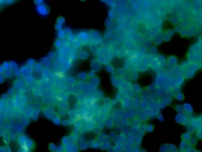
Retinal Cell Line (R28) Cytospin stained for EpCAM (green) and counterstained with DAPI (blue).
Cytospin Description: Cells are fixed for 10 minutes in cytospin solution (Contains glycerol, acetone and isopropanol). The cells are spun onto slides for 5 minutes at 1500 RPM using a Cytospin centrifuge. For details on the cytospin solution or for protocols for immunofluorescent staining and DAB staining of Cytospin slides, see Cassidy, L., et al. Journal of Biomarkers, 2013 below.
Cytospin Techniques
- Cassidy L, Choi M, Meyer J, and Seigel GM, Immunoreactivity of Pluripotent Markers SSEA-5 and L1CAM in Human Tumors, Teratomas, and Induced Pluripotent Stem Cells. Journal of Biomarkers, Volume 2013 (2013), Article ID 960862
E1A-immortalized cell characterization
- Seigel, GM, 1996. Establishment of an E1A-immortalized rat retinal cell culture, In Vitro Cell. Devel. Biol. 32: 66-68.
- Seigel, GM, Mutchler, A.L., and E.L. Imperato. 1996. Expression of glial cell markers in a retinal precursor cell line. Mol. Vis. 2: 2.
- Seigel, GM, Takahashi, M., Adamus, G., McDaniel, T. 1998. Intraocular transplantation of E1A-immortalized retinal precursor cells. Cell Transplant. 7 (6): 559-566.
- Seigel, GM, Sun, W, Wang, J., Hershberger, DH, Campbell, LM, Salvi, RJ. 2004. Neuronal gene expression and function in the growth-stimulated R28 retinal precursor cell line. Curr. Eye Res., 28 (4):257-269.
- Seigel, GM and Salvi, RJ. A microarray dataset of genes expressed by the R28 retinal precursor cell line. Dataset Papers in Neuroscience, 2013, ID: 261063. View Article
- Seigel GM. R28 retinal precursor cells: The first 20 years. Mol Vis. 2014; 20: 301306. View Article
E1A-immortalized cell utilization
- Identification of pigment epithelium-derived factor protein forms with distinct activities on tumor cell lines. Subramanian P, Deshpande M, Locatelli-Hoops S, Moghaddam-Taaheri S, Gutierrez D, Fitzgerald DP, Guerrier S, Rapp M, Notario V, Becerra SP. J Biomed Biotechnol. 2012;2012:425907. Epub 2012 Jun 4.
- Mouse acetylcholinesterase enhances neurite outgrowth of rat R28 cells through interaction with laminin-1. Sperling LE, Klaczinski J, Schütz C, Rudolph L, Layer PG. PLoS One. 2012;7(5):e36683. Epub 2012 May 3.
- Blue light stress in retinal neuronal (R28) cells is dependent on wavelength range and irradiance. Knels L, Valtink M, Roehlecke C, Lupp A, de la Vega J, Mehner M, Funk RH. Eur J Neurosci. 2011 Aug;34(4):548-58. doi: 10.1111/j.1460-9568.2011.07790.x. Epub 2011 Jul 22.
- Inhibition of reactive gliosis prevents neovascular growth in the mouse model of oxygen-induced retinopathy. DeNiro M, Al-Mohanna FH, Al-Mohanna FA. PLoS One. 2011;6(7):e22244. Epub 2011 Jul 14.
- The effects of commercially available preservative-free FDA-approved triamcinolone (Triesence®) on retinal cells in culture. Zacharias LC, Estrago-Franco MF, Ramirez C, Kenney MC, Takahashi WY, Seigel GM, Kuppermann BD. J Ocul Pharmacol Ther. 2011 Apr;27(2):143-50.
If you publish research with this product, please let us know so we can cite your paper.