Protein-Nucleic Acid Complex Crystal Screen

Currently available screens for protein-nucleic acid complex crystallization rely on the use of additives that may promote the crystallization nucleic acids dissociated from the protein. Thus, these screens can lead to false hits and wasted sample. This screen was designed specifically for protein-nucleic acid complex crystallization. The condition formulations have been developed from an in-depth analysis of actual conditions of existing protein-nucleic acid complexes in the Protein Data Bank (PDB). Through an incomplete factorial method, conditions from the nearly 2,000 deposited protein-nucleic acid complexes were summarized into an extremely efficient 48 condition screen. This screen has been successfully used by the Hollis lab for years.
- Directed screen for protein-nucleic acid complex crystallization
- Based on proven conditions from PDB
- Especially useful when amount of sample is limited
 Protein-Nucleic Acid Complex Crystal Screen Fomulation Table
Please cite the following article when publishing: Pryor EE Jr, Wozniak DJ, Hollis, T. Crystallization of Pseudomonas aeruginosa AmrZ protein: development of a comprehensive method for obtaining and optimization of protein-DNA crystals. Acta Crystallographica Section F. 2012; F68: 985-993.
From the laboratory of Thomas Hollis, PhD, Wake Forest School of Medicine.
Read Dr. Hollis's related blog post, A New Avenue for Protein-Nucleic Acid Complex Crystallization »
Currently available screens for protein-nucleic acid complex crystallization rely on the use of additives that may promote the crystallization nucleic acids dissociated from the protein. Thus, these screens can lead to false hits and wasted sample. This screen was designed specifically for protein-nucleic acid complex crystallization. The condition formulations have been developed from an in-depth analysis of actual conditions of existing protein-nucleic acid complexes in the Protein Data Bank (PDB). Through an incomplete factorial method, conditions from the nearly 2,000 deposited protein-nucleic acid complexes were summarized into an extremely efficient 48 condition screen. This screen has been successfully used by the Hollis lab for years.
- Directed screen for protein-nucleic acid complex crystallization
- Based on proven conditions from PDB
- Especially useful when amount of sample is limited
![]() Protein-Nucleic Acid Complex Crystal Screen Fomulation Table
Protein-Nucleic Acid Complex Crystal Screen Fomulation Table
Please cite the following article when publishing: Pryor EE Jr, Wozniak DJ, Hollis, T. Crystallization of Pseudomonas aeruginosa AmrZ protein: development of a comprehensive method for obtaining and optimization of protein-DNA crystals. Acta Crystallographica Section F. 2012; F68: 985-993.
From the laboratory of Thomas Hollis, PhD, Wake Forest School of Medicine.
Read Dr. Hollis's related blog post, A New Avenue for Protein-Nucleic Acid Complex Crystallization »
| Catalog Number | Product | DataSheet | Size | AVAILABILITY | Price | Qty |
|---|
| Product Type: | Kit |
| Storage: | 4C (short term), -20C (long term) |
| Shipped: | Ambient temperature |
Structures solved using Protein-Nucleic Acid Complex Crystal Screen
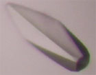 |
Transcription factor AmrZ in complex with dsDNA |
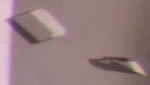 |
DNA methyl lesion repair enzyme AlkB in complex with ssDNA
PDB ID: 3KHC Holland PJ, Hollis T. Structural and mutational analysis of Escherichia coli AlkB provides insight into substrate specificity and DNA damage searching. PLoS ONE. 2010; 5(1):e8680. |
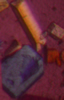 |
3' to 5' exonuclease TREX1 in complex with ssDNA
PDB ID: 2OA8 de Silva U, Choudhury S, Bailey SL, Harvey S, Perrino FW, Hollis T. The crystal structure of TREX1 explains the 3' nucleotide specificity and reveals a polyproline II helix for protein partnering. J Biol Chem. 2007; 282(14):10537-43. |
- Pryor EE Jr, Wozniak DJ, Hollis, T. Crystallization of Pseudomonas aeruginosa AmrZ protein: development of a comprehensive method for obtaining and optimization of protein-DNA crystals. Acta Crystallographica Section F. 2012; F68: 985-993.
- Pryor EE Jr, Waligora EA, Xu B, Dellos-Nolan S, Wozniak DJ, et al. The Transcription Factor AmrZ Utilizes Multiple DNA Binding Modes to Recognize Activator and Repressor Sequences of Pseudomonas aeruginosa Virulence Genes. PLoS Pathogens. 2012; 8(4): e1002648.
- Holland PJ, Hollis T. Structural and mutational analysis of Escherichia coli AlkB provides insight into substrate specificity and DNA damage searching. PLoS ONE. 2010; 5(1): e8680.
- de Silva U, Perrino FW, Hollis T. DNA binding induces active site conformational change in the human TREX2 3'-exonuclease. Nucleic Acids Res. 2009; 37(7):2411-2417.
- de Silva U, Choudhury S, Bailey SL, Harvey S, Perrino FW, Hollis T. The crystal structure of TREX1 explains the 3' nucleotide specificity and reveals a polyproline II helix for protein partnering. J Biol Chem. 2007; 282(14):10537-43
- Hollis T. Crystallization of protein-DNA complexes. In Methods Mol Biol. (ed. Doublie) 2007; 363():225-237.
- Sugiman-Marangos S, Junop M. Crystallization of the DdrB-DNA complex from Deinococcus radiodurans. Acta Crystallogr Sect F Struct Biol Cryst Commun. 2012 Dec 1;68(Pt 12):1534-7. doi: 10.1107/S1744309112044041. PubMed PMID: 23192041; PubMed Central PMCID: PMC3509982. View Article
- Choy WW, Datta D, Geiger CA, Birrane G, Grant MA. Crystallization and preliminary X-ray analysis of a complex of the FOXO1 and Ets1 DNA-binding domains and DNA. Acta Crystallogr F Struct Biol Commun. 2014 Jan;70(Pt 1):44-8. View Article
- Hadzi S, Garcia-Pino A, Gerdes K, Lah J, Loris R. Crystallization of two operator complexes from the Vibrio cholerae HigBA2 toxin-antitoxin module. Acta Crystallogr F Struct Biol Commun. 2015 Feb;71(Pt 2):226-33. View Article
- Malaby AW, Martin SK, Wood RD, Doublié S. Expression and Structural Analyses of Human DNA Polymerase θ (POLQ). Methods Enzymol. 2017;592:103-121. View Article
If you publish research with this product, please let us know so we can cite your paper.