Perkinsus marinus (CB5D4) PmMOE-GFP
 PmMOE-GFP")
P. marinus (CB5D4) PmMOE-GFP, is a mutant cell line constitutively expresses GFP (green fluorescent protein) to facilitate better understanding of parasite biology.
Perkinsus marinus is a protozoan parasite that causes “Dermo” disease (degradation of tissue) in the eastern oyster Crassostrea virginica in coastal areas of the USA. Until now, intervention strategies against the parasite have found limited success, and Dermo still remains one of the main hurdles for the restoration of oyster populations. GFP expressed in Perkinsus marinus is valuable tool to gain futhur insight in the biology of the parasite. Specifically, it will provide ample opportunities for experiments aimed at assessing gene function, as well as studies on protein targeting, interaction(s), and homologous expression/purification, among others.
From the laboratory of Gerardo R. Vasta, PhD, University of Maryland, Baltimore.
 Part of The Investigator's Annexe program.
P. marinus (CB5D4) PmMOE-GFP, is a mutant cell line constitutively expresses GFP (green fluorescent protein) to facilitate better understanding of parasite biology.
Perkinsus marinus is a protozoan parasite that causes “Dermo” disease (degradation of tissue) in the eastern oyster Crassostrea virginica in coastal areas of the USA. Until now, intervention strategies against the parasite have found limited success, and Dermo still remains one of the main hurdles for the restoration of oyster populations. GFP expressed in Perkinsus marinus is valuable tool to gain futhur insight in the biology of the parasite. Specifically, it will provide ample opportunities for experiments aimed at assessing gene function, as well as studies on protein targeting, interaction(s), and homologous expression/purification, among others.
From the laboratory of Gerardo R. Vasta, PhD, University of Maryland, Baltimore.
![]() Part of The Investigator's Annexe program.
Part of The Investigator's Annexe program.
| Product Type: | Bacteria |
| Name: | Perkinsus marinus |
| Strain: | MOE[MOE]:GFP |
| Biosafety Level: | 2 |
| Growth Conditions: | 28C, pH 6.6, 100% humidity |
| Storage: | Liquid nitrogen |
| Shipped: | Dry ice |
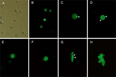
Micrographs of P. marinus trophozoites transfected with pPmMOE-GFP (with the exception of panel A, all micrographs were taken within the first weekof the transfection experiment). (A) Differential interference contrast image of trophozoites 19 h after transfection. (B) fluorescent trophozoites transfected withpPmMOE-GFP. (C, D) Trophozoites showing fluorescence in the periphery of the cell and a buildup of fluorescence in the cytoplasm, underneath the membrane(arrows). (E) Schizont containing multiple emerging fluorescent daughter cells. (F) trophozoites attached or connected to the remaining of the cell wall (arrows) afterthe rupture of the schizont. (H) Often trophozoites remained together after the schizogony. Magnifications A and B (20×) and CH (40×).
Adapted from: Fernández-Robledo JA, et al. Mol Biochem Parasitol. 2008 Jan;157(1):44-53.
- Fernández-Robledo JA, Lin Z, Vasta GR. Transfection of the protozoan parasite Perkinsus marinus. Mol Biochem Parasitol. 2008 Jan;157(1):44-53.
- Surekha Shridhar, Kolaleh Hassan, David J. Sullivan, Gerardo R. Vasta, José A. Fernández Robledo. Quantitative assessment of the proliferation of the protozoan parasite Perkinsus marinus using a bioluminescence assay for ATP content. Int J Parasitol Drugs Drug Resist. 2013 Apr 13;3:85-92.
If you publish research with this product, please let us know so we can cite your paper.