Anti-ICOS (CD278) [C398.4A] Antibody
![Anti-ICOS (CD278) [C398.4A] Antibody](https://www.kerafast.com/MediaStorage/Product/Images/Large/828_200120200126238270.jpg "Anti-ICOS (CD278) [C398.4A] Antibody")
This hamster monoclonal antibody was generated against a mouse T helper cell clone, and recognizes human, mouse, and rat ICOS.
Inducible T-cell co-stimulator (ICOS) enhances all basic T-cell responses to a foreign antigen, namely proliferation, secretion of lymphokines, up-regulation of molecules that mediate cell-cell interaction, and effective help for antibody secretion by B-cells. The protein is essential both for efficient interaction between T and B-cells and for normal antibody responses to T-cell dependent antigens. It does not up-regulate the production of interleukin-2, but superinduces the synthesis of interleukin-10. It prevents the apoptosis of pre-activated T-cells, and plays a critical role in CD40-mediated class switching of immunoglobin isotypes.
From the laboratory of Charles A. Janeway, Jr., MD, Yale University.
 Part of The Investigator's Annexe program.
This hamster monoclonal antibody was generated against a mouse T helper cell clone, and recognizes human, mouse, and rat ICOS.
Inducible T-cell co-stimulator (ICOS) enhances all basic T-cell responses to a foreign antigen, namely proliferation, secretion of lymphokines, up-regulation of molecules that mediate cell-cell interaction, and effective help for antibody secretion by B-cells. The protein is essential both for efficient interaction between T and B-cells and for normal antibody responses to T-cell dependent antigens. It does not up-regulate the production of interleukin-2, but superinduces the synthesis of interleukin-10. It prevents the apoptosis of pre-activated T-cells, and plays a critical role in CD40-mediated class switching of immunoglobin isotypes.
From the laboratory of Charles A. Janeway, Jr., MD, Yale University.
![]() Part of The Investigator's Annexe program.
Part of The Investigator's Annexe program.
| Product Type: | Antibody |
| Antigen: | Inducible T-cell co-stimulator (ICOS), CD278, AILIM, CVID1 |
| Accession ID: | Q9Y6W8 |
| Molecular Weight: | 34 kDa |
| Isotype: | Hamster Ig (unknown isotype) |
| Clonality: | Monoclonal |
| Clone Name: | C398.4A |
| Reactivity: | Mouse, rat, human |
| Immunogen: | Mouse T helper cell clone |
| Species Immunized: | Hamster |
| Purification Method: | Protein G |
| Buffer: | PBS, 0.05% (w/v) Sodium Azide |
| Tested Applications: | IP, IHC, FC |
| Storage: | -20C |
| Shipped: | Cold packs |
Immunoprecipitation
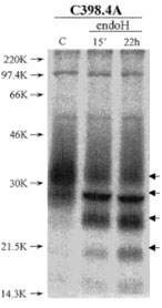
ICOS Glycosylation Patterns. Reduced immunoprecipitates of ICOS obtained from 125I-surface labeled HUT78 cell lysates were digested for the indicated times with Endo H. Lane C shows the undigested samples. Bold arrows mark the ICOS bands. Antibody C398.4A coupled to protein G-Sepharose was used. Samples were run on SDS-PAGE (14% polyacrylamide gels) under reducing conditionsand visualized with a Phospho imager.
Adapted from: Buonfiglio D, et al. Eur J Immunol. 2000 Dec;30(12):3463-7.
Immunohistochemistry

Immunohistochemical demonstration of hpH4 expression in angioimmunoblastic T cell lymphoma. Most neoplastic cells show cytoplasmic immunoreactivity for C398.4A mAb. Frozen sections; ABC method; hematoxylin counterstain. Original magnification 400 ×.
Adapted from: Buonfiglio D, et al. Eur J Immunol. 1999 Sep;29(9):2863-74.
Flow Cytometry
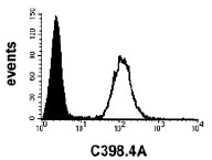
ICOS expression in L cells transfected with the human ICOS cDNA. ICOS-transfected (open curve) and wild-type cells (black curve) were stained with C398.4A followed by FITC-conjugated goat anti-mouse IgG or mouse anti-hamster IgG, respectively, and analyzed by flow cytometry.
Adapted from: Buonfiglio D, et al. Eur J Immunol. 2000 Dec;30(12):3463-7.
- Redoglia V, Dianzani U, Rojo JM, Portolés P, Bragardo M, Wolff H, Buonfiglio D, Bonissoni S, Janeway CA Jr. Characterization of H4: a mouse T lymphocyte activation molecule functionally associated with the CD3/T cell receptor. Eur J Immunol. 1996 Nov;26(11):2781-9.
- Buonfiglio D, Bragardo M, Bonissoni S, Redoglia V, Cauda R, Zupo S, Burgio VL, Wolff H, Franssila K, Gaidano G, Carbone A, Janeway CA Jr, Dianzani U. Characterization of a novel human surface molecule selectively expressed by mature thymocytes, activated T cells and subsets of T cell lymphomas. Eur J Immunol. 1999 Sep;29(9):2863-74.
- Buonfiglio D, Bragardo M, Redoglia V, Vaschetto R, Bottarel F, Bonissoni S, Bensi T, Mezzatesta C, Janeway Jr CA, Dianzani U. The T cell activation molecule H4 and the CD28-like molecule ICOS are identical. Eur J Immunol. 2000 Dec;30(12):3463-7.
If you publish research with this product, please let us know so we can cite your paper.